


1¡¢¶©¹ºÊ¹Óÿ¹Ìå²úÆ·µÄ¿Í»§£¬ÔÚʹÓòúÆ·¹ý³ÌÖÐÓöµ½ÎÊÌ⣬Ìá³ö¼¼ÊõÖ§³Ö¼°ÆäËûÇëÇóʱ£¬ÎÒ¹«Ë¾½Óµ½ÇëÇóºóµÄ24¸öСʱ֮ÄÚ×ö³ö´¦Àí¡£
2¡¢ÎÒ¹«Ë¾µÄËùÓвúÆ·¶¼¾¹ýÑϸñµÄÖʼìºóÉϼÜÏúÊÛ£¬È羸´ºËȷʵ´æÔÚÎÊÌ⣬±¾¹«Ë¾ÎÞÌõ¼þÍË¿î»ò¸ü»»»õ¡£
3¡¢ËùÓÐÊéÃæ·´À¡ÎÒÃÇÊÕµ½ºó48СʱÄÚ¸ø³ö´ð¸´¡£
| ÖÐÎÄÃû³Æ | ΢¹Üµ°°×β tubulin/Tubulin ⣨ÄڲΣ©¿¹Ìå |
| ±ð Ãû | Beta 4 tubulin; Tubulin-beta; Tubulin beta; Beta 5 tubulin; BetaTubulin; Beta-Tubulin; dJ40E16.7; TUBB; TUBB2; TUBB2A; TUBB5; tubulin beta 2A; Tubulin beta chain; Tubulin beta-5 chain; TUBB4A; TUBB4; Tubulin 5 beta; Tubulin beta-4 chain; TBB4A_HUMAN; Tubulin beta-4A chain. |
| ²úÆ·ÀàÐÍ | Äڲο¹Ìå |
| Ñо¿ÁìÓò | ÃâÒßѧ Éñ¾ÉúÎïѧ ϸ°û¹Ç¼Ü |
| ¿¹ÌåÀ´Ô´ | Rabbit |
| ¿Ë¡ÀàÐÍ | Polyclonal |
| ½»²æ·´Ó¦ | Human, Mouse, Rat, (predicted: Rabbit, ) |
| ²úÆ·Ó¦Óà | WB=1:5000-20000 ELISA=1:500-1000 IHC-P=1:100-500 IHC-F=1:100-500 Flow-Cyt=1ug/Test ICC=1:100 IF=1:100-500 £¨Ê¯À¯ÇÐƬÐè×ö¿¹ÔÐÞ¸´£© not yet tested in other applications. optimal dilutions/concentrations should be determined by the end user. |
| ·Ö ×Ó Á¿ | 55kDa |
| ϸ°û¶¨Î» | ϸ°û½¬ |
| ÐÔ ×´ | Liquid |
| Ũ ¶È | 1mg/ml |
| Ãâ Òß Ô | KLH conjugated synthetic peptide derived from human tubulin Beta:61-160/444 |
| ÑÇ ÐÍ | IgG |
| ´¿»¯·½·¨ | affinity purified by Protein A |
| ´¢ ´æ Òº | 0.01M TBS(pH7.4) with 1% BSA, 0.03% Proclin300 and 50% Glycerol. |
| ±£´æÌõ¼þ | Shipped at 4¡æ. Store at -20 °C for one year. Avoid repeated freeze/thaw cycles. |
| PubMed | PubMed |
| ²úÆ·½éÉÜ | Microtubules are constituent parts of the mitotic apparatus, cilia, flagella, and elements of the cytoskeleton. They consist principally of 2 soluble proteins, alpha- and beta-tubulin, each of about 55,000 kDa. Antibodies against beta Tubulin are useful as loading controls for Western Blotting. However it should be noted that levels of beta Tubulin may not be stable in certain cells. For example, expression of tubulin in adipose tissue is very low (Spiegelman and Farmer, Cell, 1982, 29(1):53-60) and therefore beta Tubulin should not be used as loading control for these tissues. Function: Tubulin is the major constituent of microtubules. It binds two moles of GTP, one at an exchangeable site on the beta chain and one at a non-exchangeable site on the alpha chain. Subunit: Dimer of alpha and beta chains. May interact with RNABP10 (By similarity). Interacts with PIFO. Interacts with MX1 (By similarity). Subcellular Location: Cytoplasm, cytoskeleton. Tissue Specificity: Ubiquitously expressed with highest levels in spleen, thymus and immature brain. Post-translational modifications: Some glutamate residues at the C-terminus are polyglutamylated. This modification occurs exclusively on glutamate residues and results in polyglutamate chains on the gamma-carboxyl group. Also monoglycylated but not polyglycylated due to the absence of functional TTLL10 in human. Monoglycylation is mainly limited to tubulin incorporated into axonemes (cilia and flagella) whereas glutamylation is prevalent in neuronal cells, centrioles, axonemes, and the mitotic spindle. Both modifications can coexist on the same protein on adjacent residues, and lowering glycylation levels increases polyglutamylation, and reciprocally. The precise function of such modifications is still unclear but they regulate the assembly and dynamics of axonemal microtubules (Probable). Similarity: Belongs to the tubulin family. SWISS: P07437 Gene ID: 203068 Database links: Entrez Gene: 203068 Human Omim: 191130 Human SwissProt: P07437 Human SwissProt: P99024 Mouse SwissProt: P69897 Rat
Important Note: This product as supplied is intended for research use only, not for use in human, therapeutic or diagnostic applications. ½á¹¹µ°°×£¨Structural Proteins£© tubulinÊÇÒ»ÖÖ´óÁ¿´æÔÚÓÚ²¸È鶯ÎïÄÔ×éÖ¯ÖеÄ΢¹ÜÑÇ»ùµ°°×£¬ÔڽṹÉÏÊÇÓÉÁ½¸ö¼«ÎªÏà½üµÄαºÍβÑÇ»ù×é³ÉµÄ¶þ¾ÛÌå¡¢¶à¾ÛÌåÐγÉ΢¹Üϸ˿£¬ÊÇ΢¹ÜµÄÖ÷Òª³É·Ö¡£ ΢¹Üµ°°×ÊÇÇòÐηÖ×Ó, ÓÐÁ½ÖÖÀàÐÍ:α΢¹Üµ°°×(α-tubulin)»õºÅ£ºbs-0195RºÍβ΢¹Üµ°°×(β-tubulin), ÕâÁ½ÖÖ΢¹Üµ°°×¾ßÓÐÏàËƵÄÈýά½á¹¹, Äܹ»½ôÃܵؽáºÏ³É¶þ¾ÛÌå, ×÷Ϊ΢¹Ü×é×°µÄÑÇ»ù¡£ αÑÇ»ùÓÉ450¸ö°±»ùËá×é³É, βÑÇ»ùÊÇÓÉ455¸ö°±»ùËá×é³É, ÕâÁ½ÖÖÑÇ»ùÓÐ35¡«40%µÄ°±»ùËáÐòÁÐͬԴ, ±íÃ÷±àÂëËüÃǵĻùÒò¿ÉÄÜÊÇÓÉͬһÔʼ×æÏÈÑݱä¶øÀ´. |
| ²úƷͼƬ | 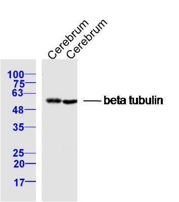 Sample: Sample:Cerebrum (Rat) Lysate at 40 ug Cerebrum (Mouse) Lysate at 40 ug Primary: Anti- Beta tubulin (bs-4511R) at 1/300 dilution Secondary: IRDye800CW Goat Anti-Rabbit IgG at 1/20000 dilution Predicted band size: 55 kD Observed band size: 55 kD 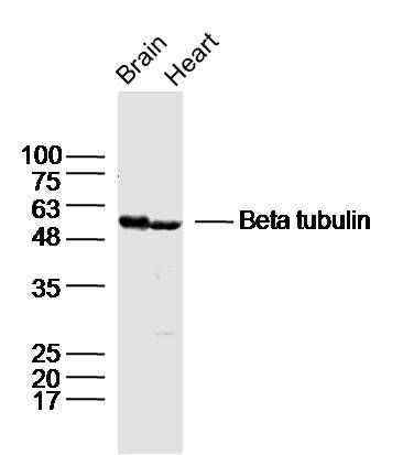 Sample: Sample:Brain (mouse) Lysate at 40 ug Heart (mouse) Lysate at 40 ug Primary: Anti- beta tubulin(bs-4511R)at 1/300 dilution Secondary: IRDye800CW Goat Anti-Rabbit IgG at 1/20000 dilution Predicted band size: 55 kD Observed band size: 55 kD 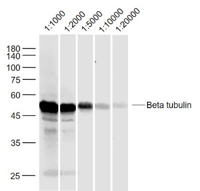 Sample: Sample:Cerebrum (Mouse) Lysate at 40 ug Primary: Anti-Beta tubulin (bs-4511R) at 1/1000~20000 dilution Secondary: IRDye800CW Goat Anti-Rabbit IgG at 1/20000 dilution Predicted band size: 55 kD Observed band size: 55 kD  Sample: Sample:Cerebrum (Rat) Lysate at 40 ug Primary: Anti-Beta tubulin (bs-4511R) at 1/1000~20000 dilution Secondary: IRDye800CW Goat Anti-Rabbit IgG at 1/20000 dilution Predicted band size: 55 kD Observed band size: 55 kD 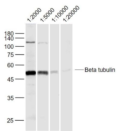 Sample: Sample:MDA-MB-231 (Human) Lysate at 40 ug Primary: Anti-Beta tubulin (bs-4511R) at 1/2000~20000 dilution Secondary: IRDye800CW Goat Anti-Rabbit IgG at 1/20000 dilution Predicted band size: 55 kD Observed band size: 55 kD 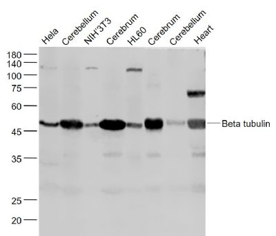 Sample: Sample:Hela(Human) Cell Lysate at 30 ug Cerebellum (Mouse) Lysate at 40 ug NIH/3T3(Mouse) Cell Lysate at 30 ug Cerebrum (Mouse) Lysate at 40 ug HL60(Human) Cell Lysate at 30 ug Cerebrum (Rat) Lysate at 40 ug Cerebellum (Rat) Lysate at 40 ug Heart (Mouse) Lysate at 40 ug Primary: Anti- Beta tubulin (bs-4511R) at 1/1000 dilution Secondary: IRDye800CW Goat Anti-Rabbit IgG at 1/20000 dilution Predicted band size: 55 kD Observed band size: 55 kD 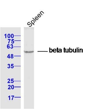 Sample: Spleen (Mouse) Lysate at 40 ug Sample: Spleen (Mouse) Lysate at 40 ugPrimary: Anti- Beta tubulin (bs-4511R) at 1/300 dilution Secondary: IRDye800CW Goat Anti-Rabbit IgG at 1/20000 dilution Predicted band size: 55 kD Observed band size: 55 kD 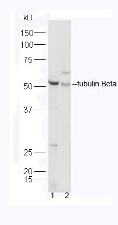 Sample: Sample:Heart (Mouse) Lysate at 40 ug MCF-7 Cell (Human) Lysate at 40 ug Primary: Anti-Beta tubulin (bs-4511R) at 1/300 dilution Secondary: HRP conjugated Goat-Anti-rabbit IgG (bs-0295G-HRP) at 1/5000 dilution Predicted band size: 55 kD Observed band size: 55 kD 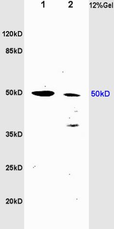 Sample: Sample:Brain (Rat) Lysate at 40 ug Heart (Rat) Lysate at 40 ug Primary: Anti-Beta tubulin (bs-4511R) at 1/300 dilution Secondary: HRP conjugated Goat-Anti-rabbit IgG (bs-0295G-HRP) at 1/5000 dilution Predicted band size: 55 kD Observed band size: 50 kD 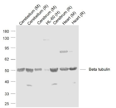 Sample: Sample:Lane 1: Cerebellum (Mouse) Lysate at 40 ug Lane 2: Cerebellum (Rat) Lysate at 40 ug Lane 3: Cerebrum (Mouse) Lysate at 40 ug Lane 4: HL-60 (Human) Cell Lysate at 30 ug Lane 5: Cerebrum (Rat) Lysate at 40 ug Lane 6: Heart (Mouse) Lysate at 40 ug Lane 7: Heart (Rat) Lysate at 40 ug Primary: Anti-Beta tubulin (bs-4511R) at 1/1000 dilution Secondary: IRDye800CW Goat Anti-Rabbit IgG at 1/20000 dilution Predicted band size: 50 kD Observed band size: 50 kD 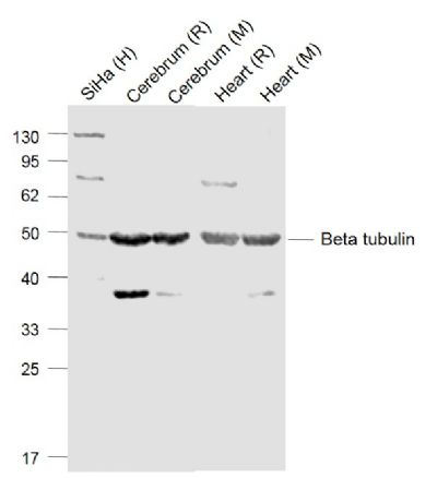 Sample: Sample:Lane 1: SiHa (Human) Cell Lysate at 30 ug Lane 2: Cerebrum (Rat) Lysate at 40 ug Lane 3: Cerebrum (Mouse) Lysate at 40 ug Lane 4: Heart (Rat) Lysate at 40 ug Lane 5: Heart (Mouse) Lysate at 40 ug Primary: Anti- Beta tubulin (bs-4511R) at 1/1000 dilution Secondary: IRDye800CW Goat Anti-Rabbit IgG at 1/20000 dilution Predicted band size: 50 kD Observed band size: 49 kD 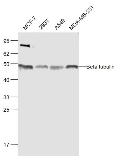 Sample: Sample:MCF-7(Human) Cell Lysate at 30 ug 293T(Human) Cell Lysate at 30 ug A549(Human) Cell Lysate at 30 ug MDA-MB-231(Human) Cell Lysate at 30 ug Primary: Anti-Beta tubulin (bs-4511R) at 1/1000 dilution Secondary: IRDye800CW Goat Anti-Rabbit IgG at 1/20000 dilution Predicted band size: 50 kD Observed band size: 48 kD 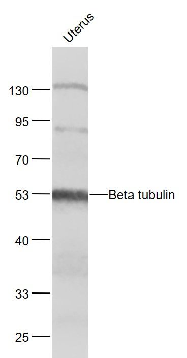 Sample: Sample:Uterus (Mouse) Lysate at 40 ug Primary: Anti- Beta tubulin (bs-4511R) at 1/1000 dilution Secondary: IRDye800CW Goat Anti-Rabbit IgG at 1/20000 dilution Predicted band size: 55 kD Observed band size: 53 kD 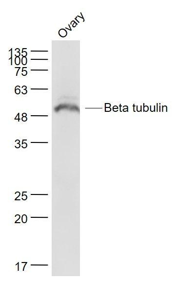 Sample: Sample:Ovary (Mouse) Lysate at 40 ug Primary: Anti- Beta tubulin (bs-4511R) at 1/1000 dilution Secondary: IRDye800CW Goat Anti-Rabbit IgG at 1/20000 dilution Predicted band size: 55 kD Observed band size: 53 kD  Paraformaldehyde-fixed, paraffin embedded (mouse cerebellum); Antigen retrieval by boiling in sodium citrate buffer (pH6.0) for 15min; Block endogenous peroxidase by 3% hydrogen peroxide for 20 minutes; Blocking buffer (normal goat serum) at 37°C for 30min; Antibody incubation with (Beta tubulin(Loading Control)) Polyclonal Antibody, Unconjugated (bs-4511R) at 1:200 overnight at 4°C, followed by operating according to SP Kit(Rabbit) (sp-0023) instructionsand DAB staining. Paraformaldehyde-fixed, paraffin embedded (mouse cerebellum); Antigen retrieval by boiling in sodium citrate buffer (pH6.0) for 15min; Block endogenous peroxidase by 3% hydrogen peroxide for 20 minutes; Blocking buffer (normal goat serum) at 37°C for 30min; Antibody incubation with (Beta tubulin(Loading Control)) Polyclonal Antibody, Unconjugated (bs-4511R) at 1:200 overnight at 4°C, followed by operating according to SP Kit(Rabbit) (sp-0023) instructionsand DAB staining. Paraformaldehyde-fixed, paraffin embedded (mouse brain); Antigen retrieval by boiling in sodium citrate buffer (pH6.0) for 15min; Block endogenous peroxidase by 3% hydrogen peroxide for 20 minutes; Blocking buffer (normal goat serum) at 37°C for 30min; Antibody incubation with (Beta tubulin(Loading Control)) Polyclonal Antibody, Unconjugated (bs-4511R) at 1:200 overnight at 4°C, followed by operating according to SP Kit(Rabbit) (sp-0023) instructionsand DAB staining. Paraformaldehyde-fixed, paraffin embedded (mouse brain); Antigen retrieval by boiling in sodium citrate buffer (pH6.0) for 15min; Block endogenous peroxidase by 3% hydrogen peroxide for 20 minutes; Blocking buffer (normal goat serum) at 37°C for 30min; Antibody incubation with (Beta tubulin(Loading Control)) Polyclonal Antibody, Unconjugated (bs-4511R) at 1:200 overnight at 4°C, followed by operating according to SP Kit(Rabbit) (sp-0023) instructionsand DAB staining. Paraformaldehyde-fixed, paraffin embedded (Mouse brain); Antigen retrieval by boiling in sodium citrate buffer (pH6.0) for 15min; Block endogenous peroxidase by 3% hydrogen peroxide for 20 minutes; Blocking buffer (normal goat serum) at 37°C for 30min; Antibody incubation with (Beta tubulin ) Polyclonal Antibody, Unconjugated (bs-4511R) at 1:400 overnight at 4°C, followed by operating according to SP Kit(Rabbit) (sp-0023) instructionsand DAB staining. Paraformaldehyde-fixed, paraffin embedded (Mouse brain); Antigen retrieval by boiling in sodium citrate buffer (pH6.0) for 15min; Block endogenous peroxidase by 3% hydrogen peroxide for 20 minutes; Blocking buffer (normal goat serum) at 37°C for 30min; Antibody incubation with (Beta tubulin ) Polyclonal Antibody, Unconjugated (bs-4511R) at 1:400 overnight at 4°C, followed by operating according to SP Kit(Rabbit) (sp-0023) instructionsand DAB staining.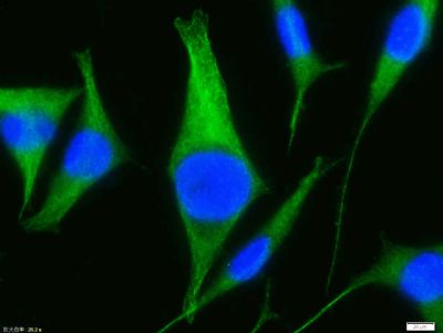 Tissue/cell:Sh-sy5y cell; 4% Paraformaldehyde-fixed; Triton X-100 at room temperature for 20 min; Blocking buffer (normal goat serum, C-0005) at 37°C for 20 min; Antibody incubation with (Beta tubulin) polyclonal Antibody, Unconjugated (bs-4511R) 1:100, 90 minutes at 37°C; followed by a FITC conjugated Goat Anti-Rabbit IgG antibody at 37°C for 90 minutes, DAPI (blue, C02-04002) was used to stain the cell nuclei. Tissue/cell:Sh-sy5y cell; 4% Paraformaldehyde-fixed; Triton X-100 at room temperature for 20 min; Blocking buffer (normal goat serum, C-0005) at 37°C for 20 min; Antibody incubation with (Beta tubulin) polyclonal Antibody, Unconjugated (bs-4511R) 1:100, 90 minutes at 37°C; followed by a FITC conjugated Goat Anti-Rabbit IgG antibody at 37°C for 90 minutes, DAPI (blue, C02-04002) was used to stain the cell nuclei.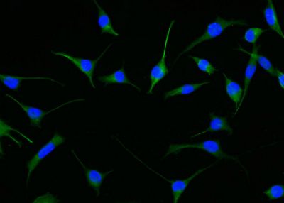 Tissue/cell:Sh-sy5y cell; 4% Paraformaldehyde-fixed; Triton X-100 at room temperature for 20 min; Blocking buffer (normal goat serum, C-0005) at 37°C for 20 min; Antibody incubation with (Beta tubulin) polyclonal Antibody, Unconjugated (bs-4511R) 1:100, 90 minutes at 37°C; followed by a FITC conjugated Goat Anti-Rabbit IgG antibody at 37°C for 90 minutes, DAPI (blue, C02-04002) was used to stain the cell nuclei. Tissue/cell:Sh-sy5y cell; 4% Paraformaldehyde-fixed; Triton X-100 at room temperature for 20 min; Blocking buffer (normal goat serum, C-0005) at 37°C for 20 min; Antibody incubation with (Beta tubulin) polyclonal Antibody, Unconjugated (bs-4511R) 1:100, 90 minutes at 37°C; followed by a FITC conjugated Goat Anti-Rabbit IgG antibody at 37°C for 90 minutes, DAPI (blue, C02-04002) was used to stain the cell nuclei. Blank control:HL-60. Blank control:HL-60.Primary Antibody (green line): Rabbit Anti-Beta tubulin antibody (bs-4511R) Dilution: 1μg /10^6 cells; Isotype Control Antibody (orange line): Rabbit IgG . Secondary Antibody : Goat anti-rabbit IgG-AF488 Dilution: 1μg /test. Protocol The cells were fixed with 4% PFA (10min at room temperature)and then permeabilized with 0.1%PBST for 20 min at room temperature. The cells were then incubated in 5%BSA to block non-specific protein-protein interactions for 30 min at room temperature .Cells stained with Primary Antibody for 30 min at room temperature. The secondary antibody used for 40 min at room temperature. Acquisition of 20,000 events was performed. |
ÎÒҪѯ¼Û
*ÁªÏµ·½Ê½£º
(¿ÉÒÔÊÇQQ¡¢MSN¡¢µç×ÓÓÊÏä¡¢µç»°µÈ£¬ÄúµÄÁªÏµ·½Ê½²»»á±»¹«¿ª)
*ÄÚÈÝ£º