


1、订购使用抗体产品的客户,在使用产品过程中遇到问题,提出技术支持及其他请求时,我公司接到请求后的24个小时之内做出处理。
2、我公司的所有产品都经过严格的质检后上架销售,如经复核确实存在问题,本公司无条件退款或更换货。
3、所有书面反馈我们收到后48小时内给出答复。
| 中文名称 | Bcl-2抗体 |
| 别 名 | Apoptosis regulator Bcl 2; Apoptosis regulator Bcl2; AW986256; B cell CLL/lymphoma 2; B cell leukemia/lymphoma 2; B cell lymphoma 2; Bcl 2; Bcl-2; Bcl2; BCL2 protein; C430015F12Rik; D630044D05Rik; D830018M01Rik; Leukemia/lymphoma, B-cell, 2; Oncogene B-cell leukemia 2; BCL2_HUMAN. |
| 研究领域 | 细胞生物 信号转导 细胞凋亡 细胞类型标志物 肿瘤细胞生物标志物 新陈代谢 线粒体 |
| 抗体来源 | Rabbit |
| 克隆类型 | Polyclonal |
| 交叉反应 | Human, (predicted: Chicken, Dog, Pig, Cow, Horse, Rabbit, Guinea Pig, ) |
| 产品应用 | WB=1:500-2000 ELISA=1:500-1000 IHC-P=1:100-500 IHC-F=1:100-500 ICC=1:100 IF=1:100-500 (石蜡切片需做抗原修复) not yet tested in other applications. optimal dilutions/concentrations should be determined by the end user. |
| 分 子 量 | 26kDa |
| 细胞定位 | 细胞核 细胞浆 细胞膜 线粒体 |
| 性 状 | Liquid |
| 浓 度 | 1mg/ml |
| 免 疫 原 | KLH conjugated synthetic peptide derived from human Bcl-2:101-160/236 |
| 亚 型 | IgG |
| 纯化方法 | affinity purified by Protein A |
| 储 存 液 | 0.01M TBS(pH7.4) with 1% BSA, 0.03% Proclin300 and 50% Glycerol. |
| 保存条件 | Shipped at 4℃. Store at -20 °C for one year. Avoid repeated freeze/thaw cycles. |
| PubMed | PubMed |
| 产品介绍 | The Bcl-2 gene was isolated at the chromosomal breakpoint of t(14;18)-bearing follicular B cell lymphomas(1,2).Bcl-2 blocks cell death following a variety of stimuli and confers a death-sparing effect to certain hematopoietic cell lines following growth factor withdrawal (3,5).Bcl-2 appears to function in several subcellular locations yet lacks any known motifs that would confer insight into its mechanism of action (6,7).A more recently identified protein,designated Bax p21(i.e., Bcl-associated X protein ),has extensive amino acid homology with Bcl-2 and both homodimerizes and forms heterodimers with Bcl-2(8). Overexpression of Bax accelerates apoptotic death induced by cytokine deprivation in an IL-3 dependent cell line and Bax also counters the death repressor activty of Bcl-2(8). Function: Suppresses apoptosis in a variety of cell systems including factor-dependent lymphohematopoietic and neural cells. Regulates cell death by controlling the mitochondrial membrane permeability. Appears to function in a feedback loop system with caspases. Inhibits caspase activity either by preventing the release of cytochrome c from the mitochondria and/or by binding to the apoptosis-activating factor (APAF-1). Subunit: Forms homodimers, and heterodimers with BAX, BAD, BAK and Bcl-X(L). Heterodimerization with BAX requires intact BH1 and BH2 motifs, and is necessary for anti-apoptotic activity. Interacts with EI24 (By similarity). Also interacts with APAF1, BBC3, BCL2L1, BNIPL, MRPL41 and TP53BP2. Binding to FKBP8 seems to target BCL2 to the mitochondria and probably interferes with the binding of BCL2 to its targets. Interacts with BAG1 in an ATP-dependent manner. Interacts with RAF1 (the 'Ser-338' and 'Ser-339' phosphorylated form). Interacts (via the BH4 domain) with EGLN3; the interaction prevents the formation of the BAX-BCL2 complex and inhibits the anti-apoptotic activity of BCL2. Interacts with G0S2; this interaction also prevents the formation of the anti-apoptotic BAX-BCL2 complex. Subcellular Location: Mitochondrion outer membrane; Single-pass membrane protein. Nucleus membrane; Single-pass membrane protein. Endoplasmic reticulum membrane; Single-pass membrane protein. Tissue Specificity: Expressed in a variety of tissues. Post-translational modifications: Phosphorylation/dephosphorylation on Ser-70 regulates anti-apoptotic activity. Growth factor-stimulated phosphorylation on Ser-70 by PKC is required for the anti-apoptosis activity and occurs during the G2/M phase of the cell cycle. In the absence of growth factors, BCL2 appears to be phosphorylated by other protein kinases such as ERKs and stress-activated kinases. Phosphorylated by MAPK8/JNK1 at Thr-69, Ser-70 and Ser-87, wich stimulates starvation-induced autophagy. Dephosphorylated by protein phosphatase 2A (PP2A). Proteolytically cleaved by caspases during apoptosis. The cleaved protein, lacking the BH4 motif, has pro-apoptotic activity, causes the release of cytochrome c into the cytosol promoting further caspase activity. Monoubiquitinated by PARK2, leading to increase its stability. DISEASE: Note=A chromosomal aberration involving BCL2 has been found in chronic lymphatic leukemia. Translocation t(14;18)(q32;q21) with immunoglobulin gene regions. BCL2 mutations found in non-Hodgkin lymphomas carrying the chromosomal translocation could be attributed to the Ig somatic hypermutation mechanism resulting in nucleotide transitions. Similarity: Belongs to the Bcl-2 family. SWISS: P49950 Gene ID: 596 Database links: Entrez Gene: 281020 Cow Entrez Gene: 596 Human Entrez Gene: 12043 Mouse Entrez Gene: 24224 Rat Omim: 151430 Human SwissProt: O02718 Cow SwissProt: P10415 Human SwissProt: P10417 Mouse SwissProt: P49950 Rat Unigene: 150749 Human Unigene: 257460 Mouse Unigene: 9996 Rat Important Note: This product as supplied is intended for research use only, not for use in human, therapeutic or diagnostic applications. Bcl-2基因是指B-cell lymphoma gene。人体滤泡B细胞淋巴瘤中过量表达的原癌基因。由于染色体t(14;18)易位,将Bcl-2基因置于免疫球蛋白重链的转录调控下,使其表达失控。在细胞系中其过量表达能延长细胞存活期而不诱导细胞增殖。它是哺乳动物中细胞调亡的抑制基因。参与细胞凋亡的调控。肿瘤中的Bcl-2基因可提高侵润性瘤细胞的生存能力。主要用于滤胞型淋巴瘤、毛细管性白血病及细胞凋亡等方面的研究。 目前研究认为:Bcl-2也是细胞凋亡的一种抑制因子、参与细胞凋亡调控,可以用于各种恶性肿瘤的细胞凋亡的研究。 |
| 产品图片 | 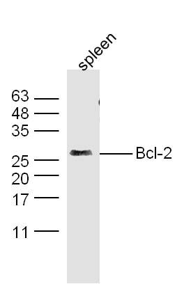 Protein: Spleen(Mouse)lysate 30ug; Protein: Spleen(Mouse)lysate 30ug;Primary: Anti-Bcl-2(bs-0032R) at 1:300; Secondary: IRDye800CW Goat Anti-Rabbit IgG at 1/10000 dilution Predicted band size : 26kD Observed band size : 26kD 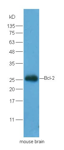 Protein: Brain(Mouse)lysate 30ug; Protein: Brain(Mouse)lysate 30ug;Primary: Anti-Bcl-2(bs-0032R) at 1:200; Secondary: HRP conjugated Goat Anti-Rabbit IgG(bs-0295G-HRP) at 1: 5000; Predicted band size : 26kD Observed band size : 26kD 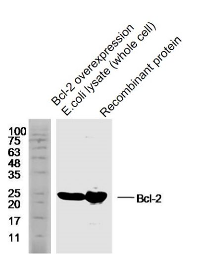 Sample: Sample:Bcl-2 overexpression E.coli lysate (whole cell) Lysate at 40 ug Recombinant protein Lysate at 40 ug Primary: Anti-Bcl-2 (bs-0032R) at 1/1000 dilution Secondary: IRDye800CW Goat Anti-Rabbit IgG at 1/20000 dilution Predicted band size: 26 kD Observed band size: 25 kD 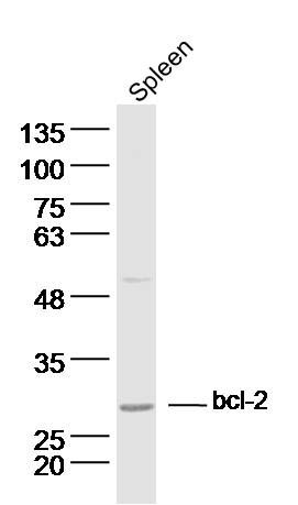 Sample:Spleen (Mouse) Lysate at 40 ug Sample:Spleen (Mouse) Lysate at 40 ugPrimary: Anti-Bcl-2 (bs-0032R) at 1/300 dilution Secondary: IRDye800CW Goat Anti-Rabbit IgG at 1/20000 dilution Predicted band size: 26 kD Observed band size: 26 kD  Sample: Sample:MCF-7 (Human) Cell Lysate at 30 ug Hela (Human) Cell Lysate at 30 ug HL60 (Human) Cell Lysate at 30 ug A431 (Human) Cell Lysate at 30 ug Primary: Anti-Bcl-2 (bs-0032R) at 1/1000 dilution Secondary: IRDye800CW Goat Anti-Rabbit IgG at 1/20000 dilution Predicted band size: 26 kD Observed band size: 23 kD 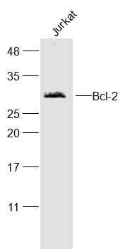 Sample: Sample:Jurkat(Human) Cell Lysate at 30 ug Primary: Anti-Bcl-2 (bs-0032R) at 1/300 dilution Secondary: IRDye800CW Goat Anti-Rabbit IgG at 1/20000 dilution Predicted band size: 26 kD Observed band size: 26 kD 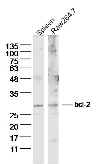 Sample: Sample:Spleen (Mouse) Lysate at 40 ug RAW264.7 Cell (Mouse) Lysate at 40 ug Primary: Anti-Bcl-2 (bs-0032R) at 1/300 dilution Secondary: IRDye800CW Goat Anti-Rabbit IgG at 1/20000 dilution Predicted band size: 26 kD Observed band size: 26 kD 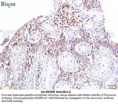   Paraformaldehyde-fixed, paraffin embedded (human pancreatic cancer); Antigen retrieval by boiling in sodium citrate buffer (pH6.0) for 15min; Block endogenous peroxidase by 3% hydrogen peroxide for 20 minutes; Blocking buffer (normal goat serum) at 37°C for 30min; Antibody incubation with (Bcl-2) Polyclonal Antibody, Unconjugated (bs-0032R) at 1:200 overnight at 4°C, followed by operating according to SP Kit(Rabbit) (sp-0023) instructionsand DAB staining. Paraformaldehyde-fixed, paraffin embedded (human pancreatic cancer); Antigen retrieval by boiling in sodium citrate buffer (pH6.0) for 15min; Block endogenous peroxidase by 3% hydrogen peroxide for 20 minutes; Blocking buffer (normal goat serum) at 37°C for 30min; Antibody incubation with (Bcl-2) Polyclonal Antibody, Unconjugated (bs-0032R) at 1:200 overnight at 4°C, followed by operating according to SP Kit(Rabbit) (sp-0023) instructionsand DAB staining. Paraformaldehyde-fixed, paraffin embedded (human pancreatic cancer); Antigen retrieval by boiling in sodium citrate buffer (pH6.0) for 15min; Block endogenous peroxidase by 3% hydrogen peroxide for 20 minutes; Blocking buffer (normal goat serum) at 37°C for 30min; Antibody incubation with (Bcl-2) Polyclonal Antibody, Unconjugated (bs-0032R) at 1:200 overnight at 4°C, followed by operating according to SP Kit(Rabbit) (sp-0023) instructionsand DAB staining. Paraformaldehyde-fixed, paraffin embedded (human pancreatic cancer); Antigen retrieval by boiling in sodium citrate buffer (pH6.0) for 15min; Block endogenous peroxidase by 3% hydrogen peroxide for 20 minutes; Blocking buffer (normal goat serum) at 37°C for 30min; Antibody incubation with (Bcl-2) Polyclonal Antibody, Unconjugated (bs-0032R) at 1:200 overnight at 4°C, followed by operating according to SP Kit(Rabbit) (sp-0023) instructionsand DAB staining. Paraformaldehyde-fixed, paraffin embedded (Rat spinal cord); Antigen retrieval by boiling in sodium citrate buffer (pH6.0) for 15min; Block endogenous peroxidase by 3% hydrogen peroxide for 20 minutes; Blocking buffer (normal goat serum) at 37°C for 30min; Antibody incubation with (Bcl-2) Polyclonal Antibody, Unconjugated (bs-0032R) at 1:400 overnight at 4°C, followed by operating according to SP Kit(Rabbit) (sp-0023) instructionsand DAB staining. Paraformaldehyde-fixed, paraffin embedded (Rat spinal cord); Antigen retrieval by boiling in sodium citrate buffer (pH6.0) for 15min; Block endogenous peroxidase by 3% hydrogen peroxide for 20 minutes; Blocking buffer (normal goat serum) at 37°C for 30min; Antibody incubation with (Bcl-2) Polyclonal Antibody, Unconjugated (bs-0032R) at 1:400 overnight at 4°C, followed by operating according to SP Kit(Rabbit) (sp-0023) instructionsand DAB staining.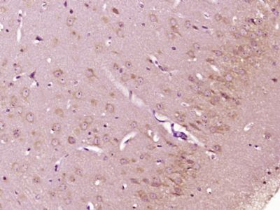 Paraformaldehyde-fixed, paraffin embedded (Mouse brain); Antigen retrieval by boiling in sodium citrate buffer (pH6.0) for 15min; Block endogenous peroxidase by 3% hydrogen peroxide for 20 minutes; Blocking buffer (normal goat serum) at 37°C for 30min; Antibody incubation with (Bcl-2) Polyclonal Antibody, Unconjugated (bs-0032R) at 1:400 overnight at 4°C, followed by operating according to SP Kit(Rabbit) (sp-0023) instructionsand DAB staining. Paraformaldehyde-fixed, paraffin embedded (Mouse brain); Antigen retrieval by boiling in sodium citrate buffer (pH6.0) for 15min; Block endogenous peroxidase by 3% hydrogen peroxide for 20 minutes; Blocking buffer (normal goat serum) at 37°C for 30min; Antibody incubation with (Bcl-2) Polyclonal Antibody, Unconjugated (bs-0032R) at 1:400 overnight at 4°C, followed by operating according to SP Kit(Rabbit) (sp-0023) instructionsand DAB staining.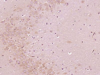 Paraformaldehyde-fixed, paraffin embedded (Rat brain); Antigen retrieval by boiling in sodium citrate buffer (pH6.0) for 15min; Block endogenous peroxidase by 3% hydrogen peroxide for 20 minutes; Blocking buffer (normal goat serum) at 37°C for 30min; Antibody incubation with (Bcl-2) Polyclonal Antibody, Unconjugated (bs-0032R) at 1:400 overnight at 4°C, followed by operating according to SP Kit(Rabbit) (sp-0023) instructionsand DAB staining. Paraformaldehyde-fixed, paraffin embedded (Rat brain); Antigen retrieval by boiling in sodium citrate buffer (pH6.0) for 15min; Block endogenous peroxidase by 3% hydrogen peroxide for 20 minutes; Blocking buffer (normal goat serum) at 37°C for 30min; Antibody incubation with (Bcl-2) Polyclonal Antibody, Unconjugated (bs-0032R) at 1:400 overnight at 4°C, followed by operating according to SP Kit(Rabbit) (sp-0023) instructionsand DAB staining.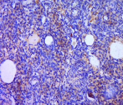 Paraformaldehyde-fixed, paraffin embedded (rat ovary tissue); Antigen retrieval by boiling in sodium citrate buffer (pH6.0) for 15min; Block endogenous peroxidase by 3% hydrogen peroxide for 20 minutes; Blocking buffer (normal goat serum) at 37°C for 30min; Antibody incubation with (Bcl-2) Polyclonal Antibody, Unconjugated (bs-0032R) at 1:400 overnight at 4°C, followed by a conjugated secondary (sp-0023) for 20 minutes and DAB staining. Paraformaldehyde-fixed, paraffin embedded (rat ovary tissue); Antigen retrieval by boiling in sodium citrate buffer (pH6.0) for 15min; Block endogenous peroxidase by 3% hydrogen peroxide for 20 minutes; Blocking buffer (normal goat serum) at 37°C for 30min; Antibody incubation with (Bcl-2) Polyclonal Antibody, Unconjugated (bs-0032R) at 1:400 overnight at 4°C, followed by a conjugated secondary (sp-0023) for 20 minutes and DAB staining. Tissue/cell: MCF-7 cell; 4% Paraformaldehyde-fixed; Triton X-100 at room temperature for 20 min; Blocking buffer (normal goat serum, C-0005) at 37°C for 20 min; Antibody incubation with (Bcl-2) Polyclonal Antibody, Unconjugated (bs-0032R) 1:50, 90 minutes at 37°C; followed by a conjugated Goat Anti-Rabbit IgG antibody (bs-0295G-Cy3) at 37°C for 90 minutes, DAPI (blue, C02-04002) was used to stain the cell nuclei. Tissue/cell: MCF-7 cell; 4% Paraformaldehyde-fixed; Triton X-100 at room temperature for 20 min; Blocking buffer (normal goat serum, C-0005) at 37°C for 20 min; Antibody incubation with (Bcl-2) Polyclonal Antibody, Unconjugated (bs-0032R) 1:50, 90 minutes at 37°C; followed by a conjugated Goat Anti-Rabbit IgG antibody (bs-0295G-Cy3) at 37°C for 90 minutes, DAPI (blue, C02-04002) was used to stain the cell nuclei. Paraformaldehyde-fixed, paraffin embedded (Rat thyroid gland); Antigen retrieval by boiling in sodium citrate buffer (pH6.0) for 15min; Block endogenous peroxidase by 3% hydrogen peroxide for 20 minutes; Blocking buffer (normal goat serum) at 37°C for 30min; Antibody incubation with (B cell lymphoma 2; Bcl-2) Polyclonal Antibody, Unconjugated (bs-0032R) at 1:200 overnight at 4°C, followed by a conjugated secondary antibody (bs-0295G-cy3) for 90 minutes and DAPI for nuclei staining. Paraformaldehyde-fixed, paraffin embedded (Rat thyroid gland); Antigen retrieval by boiling in sodium citrate buffer (pH6.0) for 15min; Block endogenous peroxidase by 3% hydrogen peroxide for 20 minutes; Blocking buffer (normal goat serum) at 37°C for 30min; Antibody incubation with (B cell lymphoma 2; Bcl-2) Polyclonal Antibody, Unconjugated (bs-0032R) at 1:200 overnight at 4°C, followed by a conjugated secondary antibody (bs-0295G-cy3) for 90 minutes and DAPI for nuclei staining. Tissue/cell: Hela cell; 4% Paraformaldehyde-fixed; Triton X-100 at room temperature for 20 min; Blocking buffer (normal goat serum, C-0005) at 37°C for 20 min; Antibody incubation with (Bcl-2) polyclonal Antibody, Unconjugated (bs-0032R) 1:100, 90 minutes at 37°C; followed by a FITC conjugated Goat Anti-Rabbit IgG antibody at 37°C for 90 minutes, DAPI (blue, C02-04002) was used to stain the cell nuclei. Tissue/cell: Hela cell; 4% Paraformaldehyde-fixed; Triton X-100 at room temperature for 20 min; Blocking buffer (normal goat serum, C-0005) at 37°C for 20 min; Antibody incubation with (Bcl-2) polyclonal Antibody, Unconjugated (bs-0032R) 1:100, 90 minutes at 37°C; followed by a FITC conjugated Goat Anti-Rabbit IgG antibody at 37°C for 90 minutes, DAPI (blue, C02-04002) was used to stain the cell nuclei. |
我要询价
*联系方式:
(可以是QQ、MSN、电子邮箱、电话等,您的联系方式不会被公开)
*内容: