


1、订购使用抗体产品的客户,在使用产品过程中遇到问题,提出技术支持及其他请求时,我公司接到请求后的24个小时之内做出处理。
2、我公司的所有产品都经过严格的质检后上架销售,如经复核确实存在问题,本公司无条件退款或更换货。
3、所有书面反馈我们收到后48小时内给出答复。
| 中文名称 | β-肌动蛋白/β-Actin(内参)抗体 |
| 别 名 | Beta Actin; beta-Actin; ACTB; Actin cytoplasmic 1; Actin, beta; Beta actin; beta cytoskeletal actin;A X actin like protein; ACTB; Actin cytoplasmic 1; alpha sarcomeric Actin; Actx; Beta cytoskeletal actin; Melanoma X actin; PS1TP5BP1; ACTB_HUMAN. |
| 产品类型 | 内参抗体 |
| 研究领域 | 肿瘤 细胞生物 信号转导 细胞骨架 |
| 抗体来源 | Rabbit |
| 克隆类型 | Polyclonal |
| 交叉反应 | Human, Mouse, Rat, mt,op (predicted: Chicken, Dog, Rabbit, Sheep, Bee, Fish, Guinea Pig, Hamster, Cat, ) |
| 产品应用 | WB=1:5000-20000 ELISA=1:5000-20000 IHC-P=1:500-1000 Flow-Cyt=1μg/Test ICC=1:100 (石蜡切片需做抗原修复) not yet tested in other applications. optimal dilutions/concentrations should be determined by the end user. |
| 分 子 量 | 42kDa |
| 细胞定位 | 细胞浆 |
| 性 状 | Liquid |
| 浓 度 | 1mg/ml |
| 免 疫 原 | Synthetic MAP peptide derived from human beta-Actin:1-200/375 |
| 亚 型 | IgG |
| 纯化方法 | affinity purified by Protein A |
| 储 存 液 | 0.01M TBS(pH7.4) with 1% BSA, 0.03% Proclin300 and 50% Glycerol. |
| 保存条件 | Shipped at 4℃. Store at -20 °C for one year. Avoid repeated freeze/thaw cycles. |
| PubMed | PubMed |
| 产品介绍 | Loading Control This gene encodes one of six different actin proteins. Actins are highly conserved proteins that are involved in cell motility, structure, and integrity. This actin is a major constituent of the contractile apparatus and one of the two nonmuscle cytoskeletal actins. [provided by RefSeq, Jul 2008]. Function: Actins are highly conserved proteins that are involved in various types of cell motility and are ubiquitously expressed in all eukaryotic cells. Subunit: Polymerization of globular actin (G-actin) leads to a structural filament (F-actin) in the form of a two-stranded helix. Each actin can bind to 4 others. Identified in a mRNP granule complex, at least composed of ACTB, ACTN4, DHX9, ERG, HNRNPA1, HNRNPA2B1, HNRNPAB, HNRNPD, HNRNPL, HNRNPR, HNRNPU, HSPA1, HSPA8, IGF2BP1, ILF2, ILF3, NCBP1, NCL, PABPC1, PABPC4, PABPN1, RPLP0, RPS3, RPS3A, RPS4X, RPS8, RPS9, SYNCRIP, TROVE2, YBX1 and untranslated mRNAs. Component of the BAF complex, which includes at least actin (ACTB), ARID1A, ARID1B/BAF250, SMARCA2, SMARCA4/BRG1, ACTL6A/BAF53, ACTL6B/BAF53B, SMARCE1/BAF57 SMARCC1/BAF155, SMARCC2/BAF170, SMARCB1/SNF5/INI1, and one or more of SMARCD1/BAF60A, SMARCD2/BAF60B, or SMARCD3/BAF60C. In muscle cells, the BAF complex also contains DPF3. Found in a complex with XPO6, Ran, ACTB and PFN1. Component of the MLL5-L complex, at least composed of MLL5, STK38, PPP1CA, PPP1CB, PPP1CC, HCFC1, ACTB and OGT. Interacts with XPO6 and EMD. Interacts with ERBB2. Subcellular Location: Cytoplasm. cytoskeleton. Tissue Specificity: Ubiquitously expressed in all eukaryotic cells. Post-translational modifications: ISGylated. Oxidation of Met-44 by MICALs (MICAL1, MICAL2 or MICAL3) to form methionine sulfoxide promotes actin filament depolymerization. Methionine sulfoxide is produced stereospecifically, but it is not known whether the (S)-S-oxide or the (R)-S-oxide is produced. DISEASE: Defects in ACTA1 are the cause of nemaline myopathy type 3 (NEM3) [MIM:161800]. A form of nemaline myopathy. Nemaline myopathies are muscular disorders characterized by muscle weakness of varying severity and onset, and abnormal thread-or rod-like structures in muscle fibers on histologic examination. The phenotype at histological level is variable. Some patients present areas devoid of oxidative activity containg (cores) within myofibers. Core lesions are unstructured and poorly circumscribed. Defects in ACTA1 are a cause of myopathy congenital with excess of thin myofilaments (MPCETM) [MIM:161800]. A congenital muscular disorder characterized at histological level by areas of sarcoplasm devoid of normal myofibrils and mitochondria, and replaced with dense masses of thin filaments. Central cores, rods, ragged red fibers, and necrosis are absent. Similarity: Belongs to the actin family. SWISS: P60709 Gene ID: 60 Database links: Entrez Gene: 396526 Chicken Entrez Gene: 60 Human Entrez Gene: 11461 Mouse Entrez Gene: 100009272 Rabbit Entrez Gene: 81822 Rat Omim: 102630 Human SwissProt: P60706 Chicken SwissProt: P60712 Cow SwissProt: P60708 Horse SwissProt: P60709 Human SwissProt: P60710 Mouse SwissProt: P29751 Rabbit SwissProt: P60711 Rat SwissProt: P60713 Sheep Unigene: 520640 Human Unigene: 708120 Human Unigene: 727576 Human Unigene: 328431 Mouse Unigene: 391967 Mouse Unigene: 94978 Rat Important Note: This product as supplied is intended for research use only, not for use in human, therapeutic or diagnostic applications. 内参抗体 β-Actin是横纹肌肌纤维中的一种主要蛋白质成分,也是肌肉细丝及细胞骨架微丝的主要成分。具有收缩功能,分布广泛,具有高度保守性,在细胞中的表达相对稳定,因此常被用作校正系统的内参。β-Actin分子量为42 kDa, 此抗体主要用于标记平滑肌及其来源的肿瘤。 我公司开发的β-Actin抗体已被国内外广大科研工作者使用,被称谓:质量信得过产品. |
| 产品图片 | 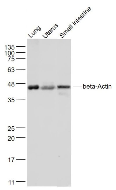 Sample: Sample:Lung (Mouse) Lysate at 40 ug Uterus (Mouse) Lysate at 40 ug Small intestine (Mouse) Lysate at 40 ug Primary: Anti- beta-Actin (bs-0061R) at 1/2000 dilution Secondary: IRDye800CW Goat Anti-Rabbit IgG at 1/20000 dilution Predicted band size: 42 kD Observed band size: 42 kD 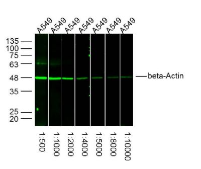 Sample: Sample:A549 Cell (Human) Lysate at 30 ug Primary: Lane1: Anti-beta-Actin (bs-0061R) at 1/500 dilution Lane2: Anti-beta-Actin (bs-0061R) at 1/1000 dilution Lane3: Anti-beta-Actin (bs-0061R) at 1/2000 dilution Lane4: Anti-beta-Actin (bs-0061R) at 1/4000 dilution Lane5: Anti-beta-Actin (bs-0061R) at 1/5000 dilution Lane6: Anti-beta-Actin (bs-0061R) at 1/8000 dilution Lane7: Anti-beta-Actin (bs-0061R) at 1/10000 dilution Secondary: IRDye800CW Goat Anti-Rabbit IgG at 1/20000 dilution Predicted band size: 42 kD Observed band size: 42 kD 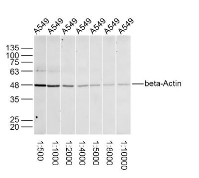 Sample: Sample:A549 Cell (Human) Lysate at 30 ug Primary: Lane1: Anti-beta-Actin (bs-0061R) at 1/500 dilution Lane2: Anti-beta-Actin (bs-0061R) at 1/1000 dilution Lane3: Anti-beta-Actin (bs-0061R) at 1/2000 dilution Lane4: Anti-beta-Actin (bs-0061R) at 1/4000 dilution Lane5: Anti-beta-Actin (bs-0061R) at 1/5000 dilution Lane6: Anti-beta-Actin (bs-0061R) at 1/8000 dilution Lane7: Anti-beta-Actin (bs-0061R) at 1/10000 dilution Secondary: IRDye800CW Goat Anti-Rabbit IgG at 1/20000 dilution Predicted band size: 42 kD Observed band size: 42 kD 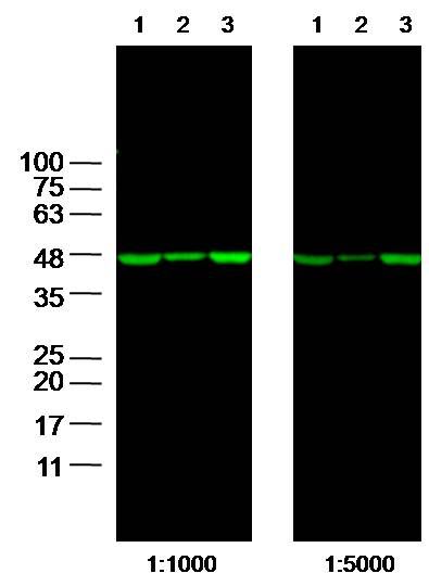 Sample: Sample:Lane1: 293T Cell Lysate at 25 ug Lane2: A549 Cell Lysate at 25 ug Lane3: A431 Cell Lysate at 25 ug Primary: Anti- beta-Actin (bs-0061R) at 1/1000 and 1/5000 dilution Secondary: IRDye800CW Goat Anti-Rabbit IgG at 1/20000 dilution Predicted band size: 42kD Observed band size: 42 kD  Sample: Lung lysate at 30ug; Sample: Lung lysate at 30ug;Primary: Anti-beta-actin (bs-0061R) at 1:1000 dilution Secondary: HRP conjugated Goat-Anti-Rabbit IgG(bse-0295G) at 1:3000 dilution Predicted band size : 42kD Observed band size : 42kD 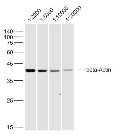 Sample: Sample:SH-SY5Y (Human) Lysate at 40 ug Primary: Anti-beta-Actin (bs-0061R) at 1/2000~1/20000 dilution Secondary: IRDye800CW Goat Anti-Rabbit IgG at 1/20000 dilution Predicted band size: 42 kD Observed band size: 42 kD 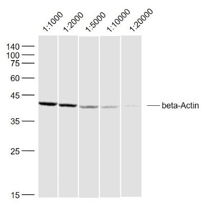 Sample: Sample:Lymph node (Rat) Lysate at 40 ug Primary: Anti-beta-Actin (bs-0061R) at 1/1000~1/20000 dilution Secondary: IRDye800CW Goat Anti-Rabbit IgG at 1/20000 dilution Predicted band size: 42 kD Observed band size: 42 kD 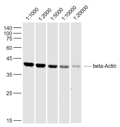 Sample: Sample:Thymus (Mouse) Lysate at 40 ug Primary: Anti-beta-Actin (bs-0061R) at 1/1000~1/20000 dilution Secondary: IRDye800CW Goat Anti-Rabbit IgG at 1/20000 dilution Predicted band size: 42 kD Observed band size: 42 kD 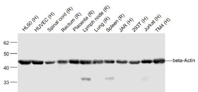 Sample: Sample:HL60 (Human) Cell Lysate at 40 ug HUVEC (Human) Cell Lysate at 40 ug Spinal cord (Rat) Lysate at 40 ug Rectum (Rat) Lysate at 40 ug Placenta (Rat) Lysate at 40 ug Lymph node (Rat) Lysate at 40 ug Lung (Rat) Lysate at 40 ug Spleen (Rat) Lysate at 40 ug JAR (Human) Cell Lysate at 40 ug 293T (Human) Cell Lysate at 40 ug Jurkat (Human) Cell Lysate at 40 ug TM4 (Human) Cell Lysate at 40 ug Primary: Anti-beta-Actin (bs-0061R) at 1/2000 dilution Secondary: IRDye800CW Goat Anti-Rabbit IgG at 1/20000 dilution Predicted band size: 42 kD Observed band size: 42 kD 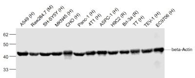 Sample: Sample:A549 (Human) Cell Lysate at 40 ug Raw264.7 (Mouse) Cell Lysate at 40 ug SH-SY5Y (Human) Cell Lysate at 40 ug MKN45 (Human) Cell Lysate at 40 ug CHO (Human) Cell Lysate at 40 ug Panc-1 (Human) Cell Lysate at 40 ug 4T1 (Human) Cell Lysate at 40 ug ASPC-1 (Human) Cell Lysate at 40 ug H9C2 (Rat) Cell Lysate at 40 ug Brl-3a (Rat) Cell Lysate at 40 ug TT (Human) Cell Lysate at 40 ug TEV-1 (Human) Cell Lysate at 40 ug EC9706 (Human) Cell Lysate at 40 ug Primary: Anti-beta-Actin (bs-0061R) at 1/2000 dilution Secondary: IRDye800CW Goat Anti-Rabbit IgG at 1/20000 dilution Predicted band size: 42 kD Observed band size: 42 kD 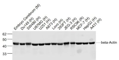 Sample: Sample:Embryo Cerebrum (Mouse) Lysate at 40 ug Du145 (Human) Lysate at 40 ug SW480 (Human) Cell Lysate at 40 ug U87MG (Human) Lysate at 40 ug U251 (Human) Lysate at 40 ug A673 (Human) Lysate at 40 ug Lovo (Human) Lysate at 40 ug 293FT (Human) Lysate at 40 ug JEG-3 (Human) Lysate at 40 ug RSC96 (Rat) Cell Lysate at 40 ug MCF-7 (Human) Cell Lysate at 40 ug HepG2 (Human) Lysate at 40 ug A431 (Human) Lysate at 40 ug Primary: Anti-beta-Actin (bs-0061R) at 1/2000 dilution Secondary: IRDye800CW Goat Anti-Rabbit IgG at 1/20000 dilution Predicted band size: 42 kD Observed band size: 42 kD 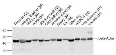 Sample: Sample:Thymus (Mouse) Lysate at 40 ug Urinary bladder (Mouse) Lysate at 40 ug Uterus (Mouse) Cell Lysate at 40 ug Aorta (Mouse) Lysate at 40 ug olfactory bulb (Mouse) Lysate at 40 ug Cerebellum (Mouse) Lysate at 40 ug Adrenal gland (Mouse) Lysate at 40 ug Ovary (Mouse) Lysate at 40 ug Ear (Mouse) Lysate at 40 ug U2os (Human) Cell Lysate at 40 ug ASPC-1 (Human) Cell Lysate at 40 ug Vas deferens (Mouse) Lysate at 40 ug trachea (Mouse) Lysate at 40 ug Primary: Anti-beta-Actin (bs-0061R) at 1/2000 dilution Secondary: IRDye800CW Goat Anti-Rabbit IgG at 1/20000 dilution Predicted band size: 42 kD Observed band size: 42 kD 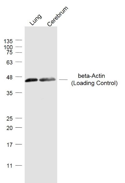 Sample: Sample:Lung (Mouse) Lysate at 40 ug Cerebrum (Mouse) Lysate at 40 ug Primary: Anti-beta-Actin (Loading Control) (bs-0061R) at 1/2000 dilution Secondary: IRDye800CW Goat Anti-Rabbit IgG at 1/20000 dilution Predicted band size: 42 kD Observed band size: 42 kD 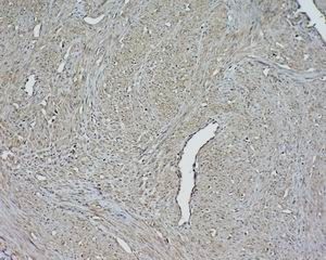 Tissue/cell: human cervical carcinoma; 4% Paraformaldehyde-fixed and paraffin-embedded; Tissue/cell: human cervical carcinoma; 4% Paraformaldehyde-fixed and paraffin-embedded;Antigen retrieval: citrate buffer ( 0.01M, pH 6.0 ), Boiling bathing for 15min; Block endogenous peroxidase by 3% Hydrogen peroxide for 30min; Blocking buffer (normal goat serum,C-0005) at 37℃ for 20 min; Incubation: Anti-Beta-actin Polyclonal Antibody, Unconjugated(bs-0061R) 1:1500, overnight at 4°C, followed by conjugation to the secondary antibody(SP-0023) and DAB(C-0010) staining 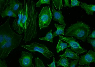 Tissue/cell: Hela cell; 4% Paraformaldehyde-fixed; Triton X-100 at room temperature for 20 min; Blocking buffer (normal goat serum, C-0005) at 37°C for 20 min; Antibody incubation with (beta-Actin) polyclonal Antibody, Unconjugated (bs-0061R) 1:100, 90 minutes at 37°C; followed by a conjugated Goat Anti-Rabbit IgG-FITC antibody at 37°C for 90 minutes, DAPI (blue, C02-04002) was used to stain the cell nuclei. Tissue/cell: Hela cell; 4% Paraformaldehyde-fixed; Triton X-100 at room temperature for 20 min; Blocking buffer (normal goat serum, C-0005) at 37°C for 20 min; Antibody incubation with (beta-Actin) polyclonal Antibody, Unconjugated (bs-0061R) 1:100, 90 minutes at 37°C; followed by a conjugated Goat Anti-Rabbit IgG-FITC antibody at 37°C for 90 minutes, DAPI (blue, C02-04002) was used to stain the cell nuclei.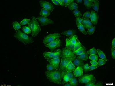 MCF7 cell; 4% Paraformaldehyde-fixed; Triton X-100 at room temperature for 20 min; Blocking buffer (normal goat serum, C-0005) at 37°C for 20 min; Antibody incubation with (beta-Actin) polyclonal Antibody, Unconjugated (bs-0061R) 1:100, 90 minutes at 37°C; followed by a conjugated Goat Anti-Rabbit IgG antibody at 37°C for 90 minutes, DAPI (blue, C02-04002) was used to stain the cell nuclei. MCF7 cell; 4% Paraformaldehyde-fixed; Triton X-100 at room temperature for 20 min; Blocking buffer (normal goat serum, C-0005) at 37°C for 20 min; Antibody incubation with (beta-Actin) polyclonal Antibody, Unconjugated (bs-0061R) 1:100, 90 minutes at 37°C; followed by a conjugated Goat Anti-Rabbit IgG antibody at 37°C for 90 minutes, DAPI (blue, C02-04002) was used to stain the cell nuclei.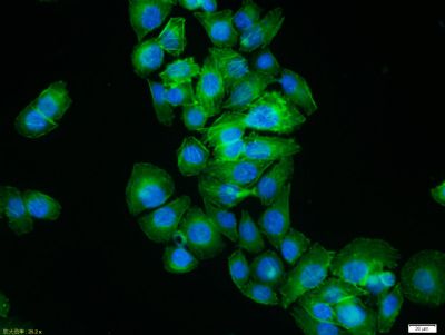 MCF7 cell; 4% Paraformaldehyde-fixed; Triton X-100 at room temperature for 20 min; Blocking buffer (normal goat serum, C-0005) at 37°C for 20 min; Antibody incubation with (beta-Actin) polyclonal Antibody, Unconjugated (bs-0061R) 1:100, 90 minutes at 37°C; followed by a conjugated Goat Anti-Rabbit IgG antibody at 37°C for 90 minutes, DAPI (blue, C02-04002) was used to stain the cell nuclei. MCF7 cell; 4% Paraformaldehyde-fixed; Triton X-100 at room temperature for 20 min; Blocking buffer (normal goat serum, C-0005) at 37°C for 20 min; Antibody incubation with (beta-Actin) polyclonal Antibody, Unconjugated (bs-0061R) 1:100, 90 minutes at 37°C; followed by a conjugated Goat Anti-Rabbit IgG antibody at 37°C for 90 minutes, DAPI (blue, C02-04002) was used to stain the cell nuclei.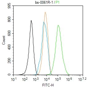 Blank control: NIH/3T3. Blank control: NIH/3T3.Primary Antibody (green line): Rabbit Anti-beta-Actin (Loading Control) antibody (bs-0061R) Dilution: 1μg /10^6 cells; Isotype Control Antibody (orange line): Rabbit IgG . Secondary Antibody : Goat anti-rabbit IgG-AF488 Dilution: 1μg /test. Protocol The cells were fixed with 4% PFA (10min at room temperature)and then permeabilized with 90% ice-cold methanol for 20 min at -20℃. The cells were then incubated in 5%BSA to block non-specific protein-protein interactions for 30 min at room temperature .Cells stained with Primary Antibody for 30 min at room temperature. The secondary antibody used for 40 min at room temperature. Acquisition of 20,000 events was performed. 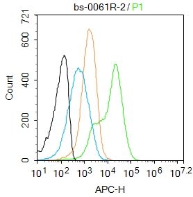 Blank control:Mouse spleen. Blank control:Mouse spleen.Primary Antibody (green line): Rabbit Anti-beta-Actin (Loading Control) antibody (bs-0061R) Dilution: 2μg /10^6 cells; Isotype Control Antibody (orange line): Rabbit IgG . Secondary Antibody : Goat anti-rabbit IgG-AF647 Dilution: 1μg /test. Protocol The cells were fixed with 4% PFA (10min at room temperature)and then permeabilized with 90% ice-cold methanol for 20 min at-20℃. The cells were then incubated in 5%BSA to block non-specific protein-protein interactions for 30 min at room temperature .Cells stained with Primary Antibody for 30 min at room temperature. The secondary antibody used for 40 min at room temperature. Acquisition of 20,000 events was performed. 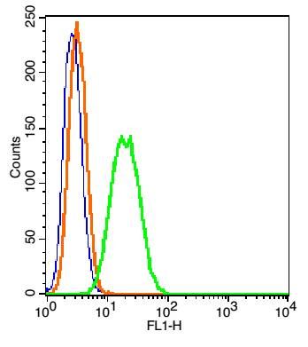 Blank control: RSC96(blue). Blank control: RSC96(blue).Primary Antibody: Rabbit Anti-beta-Actin /FITC Conjugated antibody (bs-0061R/FITC), Dilution: 1μg in 100 μL 1X PBS containing 0.5% BSA; Isotype Control Antibody: Rabbit IgG/FITC(orange) ,used under the same conditions. Protocol The cells were fixed with 2% paraformaldehyde (10 min) and then permeabilized with ice-cold 90% methanol for 30 min on ice. The cells were washed twice with 1 X PBS. The cells were incubated in 1 X PBS containing 0.5% BSA + 1 0% goat serum (15 min) to block non-specific protein-protein interactions followed by the incubated with antibody (bs-0061R/FITC, 1μg /1x10^6 cells) for 30 min on ice. Acquisition of 20,000 events was performed. |
我要询价
*联系方式:
(可以是QQ、MSN、电子邮箱、电话等,您的联系方式不会被公开)
*内容: