


1、订购使用抗体产品的客户,在使用产品过程中遇到问题,提出技术支持及其他请求时,我公司接到请求后的24个小时之内做出处理。
2、我公司的所有产品都经过严格的质检后上架销售,如经复核确实存在问题,本公司无条件退款或更换货。
3、所有书面反馈我们收到后48小时内给出答复。
| 中文名称 | 跨膜受体蛋白Notch-1抗体 |
| 别 名 | Notch 1 extracellular truncation; Notch 1 intracellular domain; hN1; Lin-12; LIN12; MIS6; Motch A; mT14; Neurogenic locus notch homolog protein 1; Neurogenic locus notch protein homolog; NICD; Notch1 intracellular domain; NOTC1_HUMAN; Notch 1; NOTCH; Notch gene homolog 1 (Drosophila); Notch homolog 1 translocation associated (Drosophila); Notch homolog 1, translocation-associated (Drosophila); NOTCH, Drosophila, homolog of, 1; notch1; p300; TAN 1; TAN1; TAN1; Translocation Associated Notch Homolog; Translocation Associated Notch Homolog; Translocation associated notch protein TAN 1; Translocation-associated notch protein TAN-1. |
| 研究领域 | 细胞生物 发育生物学 神经生物学 信号转导 干细胞 表观遗传学 |
| 抗体来源 | Rabbit |
| 克隆类型 | Polyclonal |
| 交叉反应 | Human, Mouse, Rat, |
| 产品应用 | WB=1:500-2000 ELISA=1:500-1000 IHC-P=1:100-500 IHC-F=1:100-500 Flow-Cyt=1µg/Test IF=1:100-500 (石蜡切片需做抗原修复) not yet tested in other applications. optimal dilutions/concentrations should be determined by the end user. |
| 分 子 量 | 86/89/271kDa |
| 细胞定位 | 细胞核 细胞膜 |
| 性 状 | Liquid |
| 浓 度 | 1mg/ml |
| 免 疫 原 | KLH conjugated synthetic peptide derived from human C-terminal sequence of Notch 1 extracellular truncation and Notch 1 intracellular domain :2101-2300/2555 |
| 亚 型 | IgG |
| 纯化方法 | affinity purified by Protein A |
| 储 存 液 | 0.01M TBS(pH7.4) with 1% BSA, 0.03% Proclin300 and 50% Glycerol. |
| 保存条件 | Shipped at 4℃. Store at -20 °C for one year. Avoid repeated freeze/thaw cycles. |
| PubMed | PubMed |
| 产品介绍 | This gene encodes a member of the Notch family. Members of this Type 1 transmembrane protein family share structural characteristics including an extracellular domain consisting of multiple epidermal growth factor-like (EGF) repeats, and an intracellular domain consisting of multiple, different domain types. Notch family members play a role in a variety of developmental processes by controlling cell fate decisions. The Notch signaling network is an evolutionarily conserved intercellular signaling pathway which regulates interactions between physically adjacent cells. In Drosophilia, notch interaction with its cell-bound ligands (delta, serrate) establishes an intercellular signaling pathway that plays a key role in development. Homologues of the notch-ligands have also been identified in human, but precise interactions between these ligands and the human notch homologues remain to be determined. This protein is cleaved in the trans-Golgi network, and presented on the cell surface as a heterodimer. This protein functions as a receptor for membrane bound ligands, and may play multiple roles during development. [provided by RefSeq, Jul 2008]. Function: Functions as a receptor for membrane-bound ligands Jagged1, Jagged2 and Delta1 to regulate cell-fate determination. Upon ligand activation through the released notch intracellular domain (NICD) it forms a transcriptional activator complex with RBPJ/RBPSUH and activates genes of the enhancer of split locus. Affects the implementation of differentiation, proliferation and apoptotic programs. May be important for normal lymphocyte function. In altered form, may contribute to transformation or progression in some T-cell neoplasms. Involved in the maturation of both CD4+ and CD8+ cells in the thymus. May be important for follicular differentiation and possibly cell fate selection within the follicle. During cerebellar development, may function as a receptor for neuronal DNER and may be involved in the differentiation of Bergmann glia. Subunit: Heterodimer of a C-terminal fragment N(TM) and an N-terminal fragment N(EC) which are probably linked by disulfide bonds. Interacts with DNER, DTX1, DTX2 and RBPJ/RBPSUH. Also interacts with MAML1, MAML2 and MAML3 which act as transcriptional coactivators for NOTCH1. The activated membrane-bound form interacts with AAK1 which promotes NOTCH1 stabilization. Forms a trimeric complex with FBXW7 and SGK1. Interacts with HIF1AN. HIF1AN negatively regulates the function of notch intracellular domain (NICD), accelerating myogenic differentiation. Subcellular Location: Cell membrane and Nucleus. Following proteolytical processing NICD is translocated to the nucleus. Tissue Specificity: In fetal tissues most abundant in spleen, brain stem and lung. Also present in most adult tissues where it is found mainly in lymphoid tissues. Post-translational modifications: Synthesized in the endoplasmic reticulum as an inactive form which is proteolytically cleaved by a furin-like convertase in the trans-Golgi network before it reaches the plasma membrane to yield an active, ligand-accessible form. Cleavage results in a C-terminal fragment N(TM) and a N-terminal fragment N(EC). Following ligand binding, it is cleaved by TNF-alpha converting enzyme (TACE) to yield a membrane-associated intermediate fragment called notch extracellular truncation (NEXT). This fragment is then cleaved by presenilin dependent gamma-secretase to release a notch-derived peptide containing the intracellular domain (NICD) from the membrane. Phosphorylated. O-glycosylated on the EGF-like domains. Contains both O-linked fucose and O-linked glucose. Ubiquitinated; undergoes 'Lys-29'-linked polyubiquitination catalyzed by ITCH. DISEASE: Defects in NOTCH1 are a cause of bicuspid aortic valve (BAV) [MIM:109730]. A common defect in the aortic valve in which two rather than three leaflets are present. It is often associated with aortic valve calcification and insufficiency. In extreme cases, the blood flow may be so restricted that the left ventricle fails to grow, resulting in hypoplastic left heart syndrome. Similarity: Belongs to the NOTCH family. Contains 5 ANK repeats. Contains 36 EGF-like domains. Contains 3 LNR (Lin/Notch) repeats. SWISS: P46531 Gene ID: 4851 Database links: Entrez Gene: 4851 Human Entrez Gene: 18128 Mouse Entrez Gene: 25496 Rat Omim: 190198 Human SwissProt: P46531 Human SwissProt: Q01705 Mouse SwissProt: Q07008 Rat Unigene: 495473 Human Unigene: 290610 Mouse Unigene: 25046 Rat Important Note: This product as supplied is intended for research use only, not for use in human, therapeutic or diagnostic applications. Notch 1蛋白是一个进化上保守的跨膜受体家族,广泛分布和表达在多个物种之中。研究发现Notchl作为一条信号转导途径,不仅对正常组织、细胞的分化、发育起重要作用,而且和一些肿瘤的发生和生长相关,有学者发现Notchl在许多实体瘤中异常表达,如:如宫颈癌、子宫内膜癌、肾癌、肺癌、乳腺癌、神经母细胞瘤等。因此Notchl作为一种预防和治疗肿瘤的新途径越来越受到人们的重视。 |
| 产品图片 | 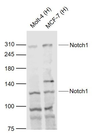 Sample: Sample:Lane 1: Molt-4 (Human) Cell Lysate at 30 ug Lane 2: MCF-7 (Human) Cell Lysate at 30 ug Primary: Anti-Notch1 (bs-1335R) at 1/1000 dilution Secondary: IRDye800CW Goat Anti-Rabbit IgG at 1/20000 dilution Predicted band size: 270/110/120 kD Observed band size: 270/110 kD  Sample: Sample:Lane 1: MCF-7 (Human) Cell Lysate at 30 ug Lane 2: A549 (Human) Cell Lysate at 30 ug Primary: Anti-Notch1 (bs-1335R) at 1/1000 dilution Secondary: IRDye800CW Goat Anti-Rabbit IgG at 1/20000 dilution Predicted band size: 270/120/110 kD Observed band size: 270/110 kD 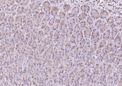 Paraformaldehyde-fixed, paraffin embedded (rat stomach); Antigen retrieval by boiling in sodium citrate buffer (pH6.0) for 15min; Block endogenous peroxidase by 3% hydrogen peroxide for 20 minutes; Blocking buffer (normal goat serum) at 37°C for 30min; Antibody incubation with (Notch1) Polyclonal Antibody, Unconjugated (bs-1335R) at 1:200 overnight at 4°C, followed by operating according to SP Kit(Rabbit) (sp-0023) instructionsand DAB staining. Paraformaldehyde-fixed, paraffin embedded (rat stomach); Antigen retrieval by boiling in sodium citrate buffer (pH6.0) for 15min; Block endogenous peroxidase by 3% hydrogen peroxide for 20 minutes; Blocking buffer (normal goat serum) at 37°C for 30min; Antibody incubation with (Notch1) Polyclonal Antibody, Unconjugated (bs-1335R) at 1:200 overnight at 4°C, followed by operating according to SP Kit(Rabbit) (sp-0023) instructionsand DAB staining.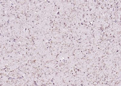 Paraformaldehyde-fixed, paraffin embedded (rat brain); Antigen retrieval by boiling in sodium citrate buffer (pH6.0) for 15min; Block endogenous peroxidase by 3% hydrogen peroxide for 20 minutes; Blocking buffer (normal goat serum) at 37°C for 30min; Antibody incubation with (Notch1) Polyclonal Antibody, Unconjugated (bs-1335R) at 1:200 overnight at 4°C, followed by operating according to SP Kit(Rabbit) (sp-0023) instructionsand DAB staining. Paraformaldehyde-fixed, paraffin embedded (rat brain); Antigen retrieval by boiling in sodium citrate buffer (pH6.0) for 15min; Block endogenous peroxidase by 3% hydrogen peroxide for 20 minutes; Blocking buffer (normal goat serum) at 37°C for 30min; Antibody incubation with (Notch1) Polyclonal Antibody, Unconjugated (bs-1335R) at 1:200 overnight at 4°C, followed by operating according to SP Kit(Rabbit) (sp-0023) instructionsand DAB staining.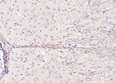 Paraformaldehyde-fixed, paraffin embedded (rat brain); Antigen retrieval by boiling in sodium citrate buffer (pH6.0) for 15min; Block endogenous peroxidase by 3% hydrogen peroxide for 20 minutes; Blocking buffer (normal goat serum) at 37°C for 30min; Antibody incubation with (Notch1) Polyclonal Antibody, Unconjugated (bs-1335R) at 1:200 overnight at 4°C, followed by operating according to SP Kit(Rabbit) (sp-0023) instructionsand DAB staining. Paraformaldehyde-fixed, paraffin embedded (rat brain); Antigen retrieval by boiling in sodium citrate buffer (pH6.0) for 15min; Block endogenous peroxidase by 3% hydrogen peroxide for 20 minutes; Blocking buffer (normal goat serum) at 37°C for 30min; Antibody incubation with (Notch1) Polyclonal Antibody, Unconjugated (bs-1335R) at 1:200 overnight at 4°C, followed by operating according to SP Kit(Rabbit) (sp-0023) instructionsand DAB staining.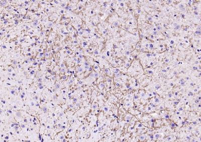 Paraformaldehyde-fixed, paraffin embedded (mouse brain); Antigen retrieval by boiling in sodium citrate buffer (pH6.0) for 15min; Block endogenous peroxidase by 3% hydrogen peroxide for 20 minutes; Blocking buffer (normal goat serum) at 37°C for 30min; Antibody incubation with (Notch1) Polyclonal Antibody, Unconjugated (bs-1335R) at 1:200 overnight at 4°C, followed by operating according to SP Kit(Rabbit) (sp-0023) instructionsand DAB staining. Paraformaldehyde-fixed, paraffin embedded (mouse brain); Antigen retrieval by boiling in sodium citrate buffer (pH6.0) for 15min; Block endogenous peroxidase by 3% hydrogen peroxide for 20 minutes; Blocking buffer (normal goat serum) at 37°C for 30min; Antibody incubation with (Notch1) Polyclonal Antibody, Unconjugated (bs-1335R) at 1:200 overnight at 4°C, followed by operating according to SP Kit(Rabbit) (sp-0023) instructionsand DAB staining.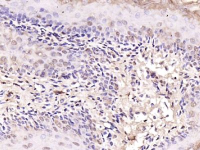 Paraformaldehyde-fixed, paraffin embedded (mouse stomach); Antigen retrieval by boiling in sodium citrate buffer (pH6.0) for 15min; Block endogenous peroxidase by 3% hydrogen peroxide for 20 minutes; Blocking buffer (normal goat serum) at 37°C for 30min; Antibody incubation with (Notch1) Polyclonal Antibody, Unconjugated (bs-1335R) at 1:200 overnight at 4°C, followed by operating according to SP Kit(Rabbit) (sp-0023) instructionsand DAB staining. Paraformaldehyde-fixed, paraffin embedded (mouse stomach); Antigen retrieval by boiling in sodium citrate buffer (pH6.0) for 15min; Block endogenous peroxidase by 3% hydrogen peroxide for 20 minutes; Blocking buffer (normal goat serum) at 37°C for 30min; Antibody incubation with (Notch1) Polyclonal Antibody, Unconjugated (bs-1335R) at 1:200 overnight at 4°C, followed by operating according to SP Kit(Rabbit) (sp-0023) instructionsand DAB staining.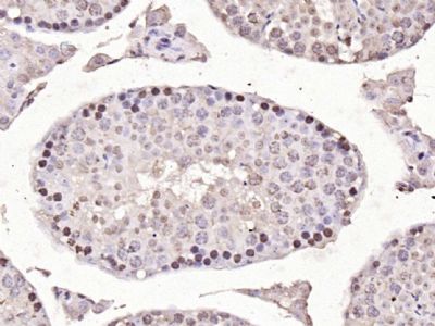 Paraformaldehyde-fixed, paraffin embedded (mouse testis); Antigen retrieval by boiling in sodium citrate buffer (pH6.0) for 15min; Block endogenous peroxidase by 3% hydrogen peroxide for 20 minutes; Blocking buffer (normal goat serum) at 37°C for 30min; Antibody incubation with (Notch1) Polyclonal Antibody, Unconjugated (bs-1335R) at 1:200 overnight at 4°C, followed by operating according to SP Kit(Rabbit) (sp-0023) instructionsand DAB staining. Paraformaldehyde-fixed, paraffin embedded (mouse testis); Antigen retrieval by boiling in sodium citrate buffer (pH6.0) for 15min; Block endogenous peroxidase by 3% hydrogen peroxide for 20 minutes; Blocking buffer (normal goat serum) at 37°C for 30min; Antibody incubation with (Notch1) Polyclonal Antibody, Unconjugated (bs-1335R) at 1:200 overnight at 4°C, followed by operating according to SP Kit(Rabbit) (sp-0023) instructionsand DAB staining. Paraformaldehyde-fixed, paraffin embedded (rat stomach); Antigen retrieval by boiling in sodium citrate buffer (pH6.0) for 15min; Block endogenous peroxidase by 3% hydrogen peroxide for 20 minutes; Blocking buffer (normal goat serum) at 37°C for 30min; Antibody incubation with (Notch1) Polyclonal Antibody, Unconjugated (bs-1335R) at 1:200 overnight at 4°C, followed by operating according to SP Kit(Rabbit) (sp-0023) instructionsand DAB staining. Paraformaldehyde-fixed, paraffin embedded (rat stomach); Antigen retrieval by boiling in sodium citrate buffer (pH6.0) for 15min; Block endogenous peroxidase by 3% hydrogen peroxide for 20 minutes; Blocking buffer (normal goat serum) at 37°C for 30min; Antibody incubation with (Notch1) Polyclonal Antibody, Unconjugated (bs-1335R) at 1:200 overnight at 4°C, followed by operating according to SP Kit(Rabbit) (sp-0023) instructionsand DAB staining.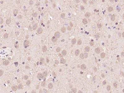 Paraformaldehyde-fixed, paraffin embedded (rat brain); Antigen retrieval by boiling in sodium citrate buffer (pH6.0) for 15min; Block endogenous peroxidase by 3% hydrogen peroxide for 20 minutes; Blocking buffer (normal goat serum) at 37°C for 30min; Antibody incubation with (Notch1) Polyclonal Antibody, Unconjugated (bs-1335R) at 1:200 overnight at 4°C, followed by operating according to SP Kit(Rabbit) (sp-0023) instructionsand DAB staining. Paraformaldehyde-fixed, paraffin embedded (rat brain); Antigen retrieval by boiling in sodium citrate buffer (pH6.0) for 15min; Block endogenous peroxidase by 3% hydrogen peroxide for 20 minutes; Blocking buffer (normal goat serum) at 37°C for 30min; Antibody incubation with (Notch1) Polyclonal Antibody, Unconjugated (bs-1335R) at 1:200 overnight at 4°C, followed by operating according to SP Kit(Rabbit) (sp-0023) instructionsand DAB staining.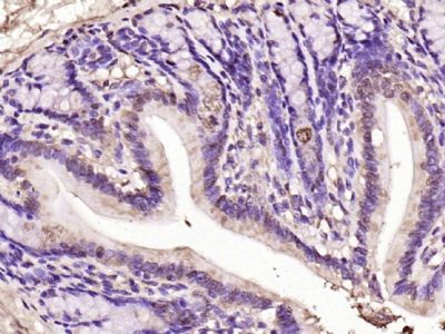 Paraformaldehyde-fixed, paraffin embedded (rat colon); Antigen retrieval by boiling in sodium citrate buffer (pH6.0) for 15min; Block endogenous peroxidase by 3% hydrogen peroxide for 20 minutes; Blocking buffer (normal goat serum) at 37°C for 30min; Antibody incubation with (Notch1) Polyclonal Antibody, Unconjugated (bs-1335R) at 1:200 overnight at 4°C, followed by operating according to SP Kit(Rabbit) (sp-0023) instructionsand DAB staining. Paraformaldehyde-fixed, paraffin embedded (rat colon); Antigen retrieval by boiling in sodium citrate buffer (pH6.0) for 15min; Block endogenous peroxidase by 3% hydrogen peroxide for 20 minutes; Blocking buffer (normal goat serum) at 37°C for 30min; Antibody incubation with (Notch1) Polyclonal Antibody, Unconjugated (bs-1335R) at 1:200 overnight at 4°C, followed by operating according to SP Kit(Rabbit) (sp-0023) instructionsand DAB staining.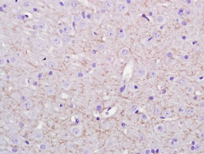 Paraformaldehyde-fixed, paraffin embedded (Rat brain); Antigen retrieval by boiling in sodium citrate buffer (pH6.0) for 15min; Block endogenous peroxidase by 3% hydrogen peroxide for 20 minutes; Blocking buffer (normal goat serum) at 37°C for 30min; Antibody incubation with (Notch1) Polyclonal Antibody, Unconjugated (bs-1335R) at 1:400 overnight at 4°C, followed by a conjugated secondary antibody (sp-0023) for 20 minutes and DAB staining. Paraformaldehyde-fixed, paraffin embedded (Rat brain); Antigen retrieval by boiling in sodium citrate buffer (pH6.0) for 15min; Block endogenous peroxidase by 3% hydrogen peroxide for 20 minutes; Blocking buffer (normal goat serum) at 37°C for 30min; Antibody incubation with (Notch1) Polyclonal Antibody, Unconjugated (bs-1335R) at 1:400 overnight at 4°C, followed by a conjugated secondary antibody (sp-0023) for 20 minutes and DAB staining.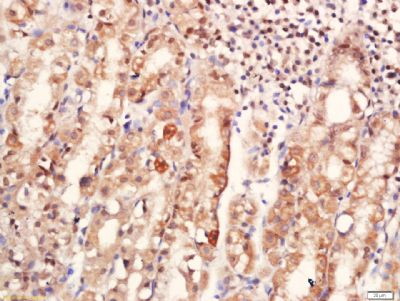 Tissue/cell: human stomach tissue; 4% Paraformaldehyde-fixed and paraffin-embedded; Tissue/cell: human stomach tissue; 4% Paraformaldehyde-fixed and paraffin-embedded;Antigen retrieval: citrate buffer ( 0.01M, pH 6.0 ), Boiling bathing for 15min; Block endogenous peroxidase by 3% Hydrogen peroxide for 30min; Blocking buffer (normal goat serum,C-0005) at 37℃ for 20 min; Incubation: Anti-Notch1 Polyclonal Antibody, Unconjugated(bs-1335R) 1:500, overnight at 4°C, followed by conjugation to the secondary antibody(SP-0023) and DAB(C-0010) staining 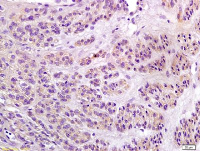 Tissue/cell: Human endometrium carcinoma; 4% Paraformaldehyde-fixed and paraffin-embedded; Tissue/cell: Human endometrium carcinoma; 4% Paraformaldehyde-fixed and paraffin-embedded;Antigen retrieval: citrate buffer ( 0.01M, pH 6.0 ), Boiling bathing for 15min; Block endogenous peroxidase by 3% Hydrogen peroxide for 30min; Blocking buffer (normal goat serum,C-0005) at 37℃ for 20 min; Incubation: Anti-Notch1 Polyclonal Antibody, Unconjugated(bs-1335R) 1:200, overnight at 4°C, followed by conjugation to the secondary antibody(SP-0023) and DAB(C-0010) staining 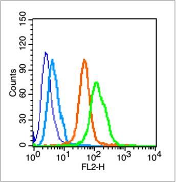 Blank control (blue line): Hela (blue). Blank control (blue line): Hela (blue).Primary Antibody (green line): Rabbit Anti-Notch1 antibody (bs-1335R) Dilution: 1μg /10^6 cells; Isotype Control Antibody (orange line): Rabbit IgG . Secondary Antibody (white blue line): Goat anti-rabbit IgG-PE Dilution: 1μg /test. Protocol The cells were fixed with 70% ethanol overnight at 4℃ and then permeabilized with 90% ice-cold methanol for 30 min on ice. Cells stained with Primary Antibody for 30 min at room temperature. The cells were then incubated in 1 X PBS/2%BSA/10% goat serum to block non-specific protein-protein interactions followed by the antibody for 15 min at room temperature. The secondary antibody used for 40 min at room temperature. Acquisition of 20,000 events was performed. |
我要询价
*联系方式:
(可以是QQ、MSN、电子邮箱、电话等,您的联系方式不会被公开)
*内容: