

1¡¢¶©¹ºÊ¹Óÿ¹Ìå²úÆ·µÄ¿Í»§£¬ÔÚʹÓòúÆ·¹ý³ÌÖÐÓöµ½ÎÊÌ⣬Ìá³ö¼¼ÊõÖ§³Ö¼°ÆäËûÇëÇóʱ£¬ÎÒ¹«Ë¾½Óµ½ÇëÇóºóµÄ24¸öСʱ֮ÄÚ×ö³ö´¦Àí¡£
2¡¢ÎÒ¹«Ë¾µÄËùÓвúÆ·¶¼¾¹ýÑϸñµÄÖʼìºóÉϼÜÏúÊÛ£¬È羸´ºËȷʵ´æÔÚÎÊÌ⣬±¾¹«Ë¾ÎÞÌõ¼þÍË¿î»ò¸ü»»»õ¡£
3¡¢ËùÓÐÊéÃæ·´À¡ÎÒÃÇÊÕµ½ºó48СʱÄÚ¸ø³ö´ð¸´¡£
| ÖÐÎÄÃû³Æ | BºÍTÁÜ°Íϸ°ûË¥¼õµ°°×¿¹Ìå |
| ±ð Ãû | B and T lymphocyte associated protein; B and T lymphocyte attenuator; B and T lymphocyte associated; BTLA; BTLA1; CD272 antigen; FLJ16065; MGC129743; BTLA_HUMAN; B- and T-lymphocyte attenuator; B- and T-lymphocyte-associated protein; CD272. |
| Ñо¿ÁìÓò | Ö×Áö ϸ°ûÉúÎï ÃâÒßѧ ÁÜ°Íϸ°û t-ÁÜ°Íϸ°û b-ÁÜ°Íϸ°û |
| ¿¹ÌåÀ´Ô´ | Rabbit |
| ¿Ë¡ÀàÐÍ | Polyclonal |
| ½»²æ·´Ó¦ | Human, Mouse, (predicted: Rat, ) |
| ²úÆ·Ó¦Óà | WB=1:500-2000 ELISA=1:500-1000 IHC-P=1:100-500 IHC-F=1:100-500 Flow-Cyt=1μg /test ICC=1:100-500 IF=1:100-500 £¨Ê¯À¯ÇÐƬÐè×ö¿¹ÔÐÞ¸´£© not yet tested in other applications. optimal dilutions/concentrations should be determined by the end user. |
| ·Ö ×Ó Á¿ | 28kDa |
| ϸ°û¶¨Î» | ϸ°ûĤ |
| ÐÔ ×´ | Liquid |
| Ũ ¶È | 1mg/ml |
| Ãâ Òß Ô | KLH conjugated synthetic peptide derived from human B and T-lymphocyte attenuator:221-289/289 |
| ÑÇ ÐÍ | IgG |
| ´¿»¯·½·¨ | affinity purified by Protein A |
| ´¢ ´æ Òº | 0.01M TBS(pH7.4) with 1% BSA, 0.03% Proclin300 and 50% Glycerol. |
| ±£´æÌõ¼þ | Shipped at 4¡æ. Store at -20 °C for one year. Avoid repeated freeze/thaw cycles. |
| PubMed | PubMed |
| ²úÆ·½éÉÜ | B and T lymphocyte attenuator (BTLA), an immunoglobulin domain-containing glycoprotein with two immunoreceptor tyrosine-based inhibitory motifs. BTLA is not expressed by naive T cells, but it is induced during activation and remains expressed on T helper type 1 (T(H)1) but not T(H)2 cells. Crosslinking BTLA with antigen receptors induces its tyrosine phosphorylation and association with the Src homology domain 2 (SH2)-containing protein tyrosine phosphatases SHP-1 and SHP-2, and attenuates production of interleukin 2 (IL-2). BTLA-deficient T cells show increased proliferation, and BTLA-deficient mice have increased specific antibody responses and enhanced sensitivity to experimental autoimmune encephalomyelitis. B7x, a peripheral homolog of B7, is a ligand of BTLA. Thus, BTLA is a third inhibitory receptor on T lymphocytes with similarities to cytotoxic T lymphocyte-associated antigen 4 (CTLA-4) and programmed death 1 (PD-1). Function: Lymphocyte inhibitory receptor which inhibits lymphocytes during immune response. Subunit: Interacts with tyrosine phosphatases PTPN6/SHP-1 and PTPN11/SHP-2. Interacts with TNFRSF14/HVEM. Subcellular Location: Membrane; Single-pass type I membrane protein (Potential). Post-translational modifications: Phosphorylated on Tyr residues by TNFRSF14 and by antigen receptors cross-linking, both inducing association with PTPN6 and PTPN11. N-glycosylated. Similarity: Contains 1 Ig-like V-type (immunoglobulin-like) domain. SWISS: Q7Z6A9 Gene ID: 151888 Database links: Entrez Gene: 151888 Human Entrez Gene: 208154 Mouse Entrez Gene: 407756 Rat Omim: 607925 Human SwissProt: Q7Z6A9 Human SwissProt: Q7TSA3 Mouse SwissProt: Q6PNM1 Rat Unigene: 445162 Human Unigene: 38199 Mouse Unigene: 124474 Rat Important Note: This product as supplied is intended for research use only, not for use in human, therapeutic or diagnostic applications. BTLA¶ÔTϸ°ûµÄ»î»¯¡¢ÔöÖ³Æð×ÅÖØÒªµÄ¸ºµ÷¿Ø×÷Óã¬BTLAÏàÓ¦ÅäÌåΪTNFR³¬¼Ò×åÖеÄðåÕ¶¾ÈëÇÖ½éÖÊ£¨HVEM£©, Æä±í´ïÓÚ°üÀ¨Tϸ°ûÔÚÄڵĶàÖÖÃâÒßϸ°û±íÃæ¡£ ÓÐѧÕß½«Ëû¶¨ÎªCD28µÄ³¬¼¶×å³ÉÔ±£¬B¡¢TÁÜ°Íϸ°ûË¥¼õ×Ó-CD272Ö÷ÒªÓÃÓÚϸ°ûÐźŴ«µ¼·½ÃæµÄÑо¿¡£ ½üÀ´¹úÍâѧÕ߶ÔBTLAÓÃÓÚÒÖÖÆÖ×Áö·½ÃæµÄÑо¿Ò²ÓÐÁËеĽøÕ¹£¬ÈÏΪB¡¢TÁÜ°Íϸ°ûË¥¼õ×Ó¶ÔÖ×ÁöµÄÉú³¤ÓÐÒÖÖÆ×÷Óã¬Ì½Ë÷еÄÖ×ÁöÃâÒßÖÎÁÆÓÐÁËеÄ;¾¶£¬ ·â±Õ´Ë;¾¶ÓпÉÄܳÉΪÖ×ÁöÃâÒßÖÎÁƵÄае㡣 |
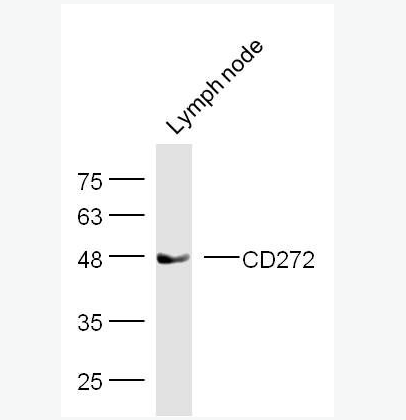
| ²úƷͼƬ | 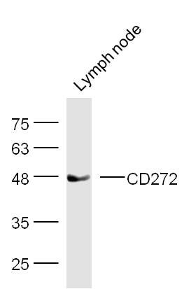 Sample: Lymph node (Mouse) Lysate at 40 ug Sample: Lymph node (Mouse) Lysate at 40 ugPrimary: Anti-CD272 (bs-0624R) at 1/300 dilution Secondary: IRDye800CW Goat Anti-Rabbit IgG at 1/10000 dilution Predicted band size: 28 kD Observed band size: 48 kD 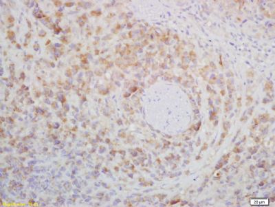 Tissue/cell: mouse lymphoma tissue; 4% Paraformaldehyde-fixed and paraffin-embedded; Tissue/cell: mouse lymphoma tissue; 4% Paraformaldehyde-fixed and paraffin-embedded;Antigen retrieval: citrate buffer ( 0.01M, pH 6.0 ), Boiling bathing for 15min; Block endogenous peroxidase by 3% Hydrogen peroxide for 30min; Blocking buffer (normal goat serum,C-0005) at 37∩ for 20 min; Incubation: Anti-CD272/BTLA Polyclonal Antibody, Unconjugated(bs-0624R) 1:200, overnight at 4∑C, followed by conjugation to the secondary antibody(SP-0023) and DAB(C-0010) staining 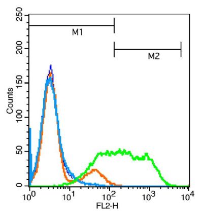 Blank control: Jurkat cells(blue). Blank control: Jurkat cells(blue).Primary Antibody:Rabbit Anti- CD272 antibody(bs-0624R), Dilution: 1μg in 100 μL 1X PBS containing 0.5% BSA; Isotype Control Antibody: Rabbit IgG(orange) ,used under the same conditions ); Secondary Antibody: Goat anti-rabbit IgG-PE(white blue), Dilution: 1:200 in 1 X PBS containing 0.5% BSA. Protocol The cells were fixed with 2% paraformaldehyde (10 min) . Primary antibody (bs-0624R, 1μg /1x10^6 cells) were incubated for 30 min on the ice, followed by 1 X PBS containing 0.5% BSA + 1 0% goat serum (15 min) to block non-specific protein-protein interactions. Then the Goat Anti-rabbit IgG/PE antibody was added into the blocking buffer mentioned above to react with the primary antibody at 1/200 dilution for 30 min on ice. Acquisition of 20,000 events was performed. |
ÎÒҪѯ¼Û
*ÁªÏµ·½Ê½£º
(¿ÉÒÔÊÇQQ¡¢MSN¡¢µç×ÓÓÊÏä¡¢µç»°µÈ£¬ÄúµÄÁªÏµ·½Ê½²»»á±»¹«¿ª)
*ÄÚÈÝ£º