

1”¢¶©¹ŗŹ¹ÓĆæ¹Ģå²śĘ·µÄæĶ»§£¬ŌŚŹ¹ÓĆ²śĘ·¹ż³ĢÖŠÓöµ½ĪŹĢā£¬Ģį³ö¼¼ŹõÖ§³Ö¼°ĘäĖūĒėĒóŹ±£¬ĪŅ¹«Ė¾½Óµ½ĒėĒóŗóµÄ24øöŠ”Ź±Ö®ÄŚ×ö³ö“¦Ąķ”£
2”¢ĪŅ¹«Ė¾µÄĖłÓŠ²śĘ·¶¼¾¹żŃĻøńµÄÖŹ¼ģŗóÉĻ¼ÜĻśŹŪ£¬Čē¾ø“ŗĖČ·Źµ“ęŌŚĪŹĢā£¬±¾¹«Ė¾ĪŽĢõ¼žĶĖæī»ņøü»»»õ”£
3”¢ĖłÓŠŹéĆę·“Ą”ĪŅĆĒŹÕµ½ŗó48Š”Ź±ÄŚøų³ö“šø“”£
Ó¢ĪÄĆū³ĘGFAP
ÖŠĪÄĆū³Ę½ŗÖŹĻĖĪ¬ĖįŠŌµ°°×µ„æĖĀ”æ¹Ģå
±š ĆūAstrocyte; FLJ45472; GFAP; Glial Fibrillary Acidic Protein; Intermediate filament protein; GFAP_HUMAN.
ŃŠ¾æĮģÓņÖ×Įö Ļø°ūÉśĪļ Éń¾ÉśĪļѧ
æ¹ĢåĄ“Ō“Mouse
æĖĀ”ĄąŠĶMonoclonal
æĖ Ā” ŗÅ7D8
½»²ę·“Ó¦Mouse, Rat,
²śĘ·Ó¦ÓĆWB=1:500-1000 IHC-P=1:100-500 IHC-F=1:100-500 ICC=1:100-500 £ØŹÆĄÆĒŠĘ¬Šč×öæ¹ŌŠŽø“£©
not yet tested in other applications.
optimal dilutions/concentrations should be determined by the end user.
·Ö ×Ó Įæ49kDa
Ļø°ū¶ØĪ»Ļø°ū½¬
ŠŌ דLiquid
ÅØ ¶Č1mg/ml
Ćā Ņß ŌRecombinant mouse GFAP full length:
ŃĒ ŠĶIgG
“æ»Æ·½·Øaffinity purified by Protein G
“¢ “ę Ņŗ0.01M TBS(pH7.4) with 1% BSA, 0.03% Proclin300 and 50% Glycerol.
±£“ęĢõ¼žShipped at 4”ę. Store at -20 °C for one year. Avoid repeated freeze/thaw cycles.
PubMedPubMed
²śĘ·½éÉÜThis gene encodes one of the major intermediate filament proteins of mature astrocytes. It is used as a marker to distinguish astrocytes from other glial cells during development. Mutations in this gene cause Alexander disease, a rare disorder of astrocytes in the central nervous system. Alternative splicing results in multiple transcript variants encoding distinct isoforms. [provided by RefSeq, Oct 2008]
Function:
GFAP, a class-III intermediate filament, is a cell-specific marker that, during the development of the central nervous system, distinguishes astrocytes from other glial cells.
Subunit:
Interacts with SYNM. Isoform 3 interacts with PSEN1 (via N-terminus).
Subcellular Location:
Cytoplasm. Note=Associated with intermediate filaments.
Tissue Specificity:
Expressed in cells lacking fibronectin.
Post-translational modifications:
Phosphorylated by PKN1.
DISEASE:
Defects in GFAP are a cause of Alexander disease (ALEXD) [MIM:203450]. Alexander disease is a rare disorder of the central nervous system. It is a progressive leukoencephalopathy whose hallmark is the widespread accumulation of Rosenthal fibers which are cytoplasmic inclusions in astrocytes. The most common form affects infants and young children, and is characterized by progressive failure of central myelination, usually leading to death usually within the first decade. Infants with Alexander disease develop a leukoencephalopathy with macrocephaly, seizures, and psychomotor retardation. Patients with juvenile or adult forms typically experience ataxia, bulbar signs and spasticity, and a more slowly progressive course.
Similarity:
Belongs to the intermediate filament family.
SWISS:
P14136
Gene ID:
2670
Database links:
Entrez Gene: 281189 Cow
Entrez Gene: 2670 Human
Entrez Gene: 14580 Mouse
Entrez Gene: 24387 Rat
Omim: 137780 Human
SwissProt: Q28115 Cow
SwissProt: P14136 Human
SwissProt: P03995 Mouse
Important Note:
This product as supplied is intended for research use only, not for use in human, therapeutic or diagnostic applications.
ŠĒŠĪ½ŗÖŹĻø°ū±źÖ¾Īļ £ØAstrocyte Marker£©
GFAPŹĒŅ»øö56kDaµÄÖŠ¼äĖæµ°°×£Øintermediate filament£¬IF£©£¬ŌŚÖŠŹąÉń¾ĻµĶ³·¢ÓżĘŚŹĒŅ»øöĢŲŅģŠŌµÄ±źÖ¾Īļ£¬ŅŌĒų±šŠĒŠĪĻø°ūŗĶĘäĖü½ŗÖŹĻø°ū”£GFAP±ķ“ļŌŚĘ¤²ćŗĶŗ£Āķ,¼±”¢ĀżŠŌʤ֏ĶŖÖĪĮĘŹ±±ķ“ļ¼õÉŁ”£
GFAPæÉŅŌŗĶČĖ”¢“óŹó”¢Š”ŹóµÄGFAP·“Ó¦£¬ŌŚÕż³£ŗĶÖ×ĮöŠŌµÄŠĒŠĪ½ŗÖŹĻø°ūŃōŠŌ±ķ“ļ£¬¶ųÉń¾½ŚĻø°ū”¢Éń¾ŌŖ”¢³ÉĻĖĪ¬Ļø°ū”¢ÉŁĶ»½ŗÖŹĻø°ūŗĶÕāŠ©Ļø°ūĄ“Ō“µÄÖ×ĮöĻø°ūŅõŠŌ±ķ“ļ£¬Ö÷ŅŖÓĆÓŚŠĒŠĪ½ŗÖŹĮöµČÖŠŹąÉń¾ĻµĶ³Ö×ĮöµÄÕļ¶ĻŗĶ¼ų±šÕļ¶Ļ,GFAPµÄȱ·¦æɵ¼ÖĀAD²””£
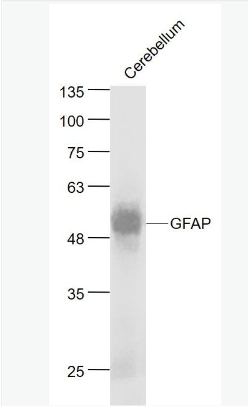
| ²śĘ·Ķ¼Ę¬ | 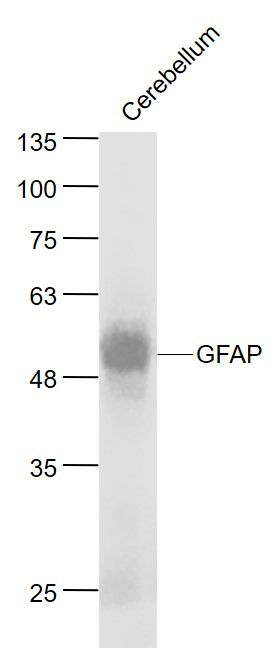 Sample: Cerebellum (Mouse) Lysate at 40 ug Primary: Anti- GFAP (bsm-33065M) at 1/1000 dilution Secondary: IRDye800CW Goat Anti-Rabbit IgG at 1/20000 dilution Predicted band size: 50 kD Observed band size: 50 kD 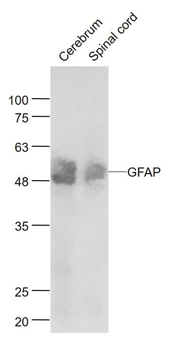 Sample: Cerebrum (Mouse) Lysate at 40 ug Spinal cord (Mouse) Lysate at 40 ug Primary: Anti- TBX1 (bsm-33065M) at 1/1000 dilution Secondary: IRDye800CW Goat Anti-Rabbit IgG at 1/20000 dilution Predicted band size: 50 kD Observed band size: 50 kD 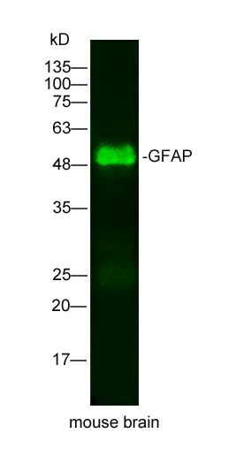 Sample: mouse brain Lysate at 25 ug Primary: Mouse Anti-GFAP(bsm-33065M) at 1/500 dilution Secondary: IRDye800CW Goat Anti-Mouse IgG at 1/20000 dilution Predicted band size: 49kD Observed band size: 49kD 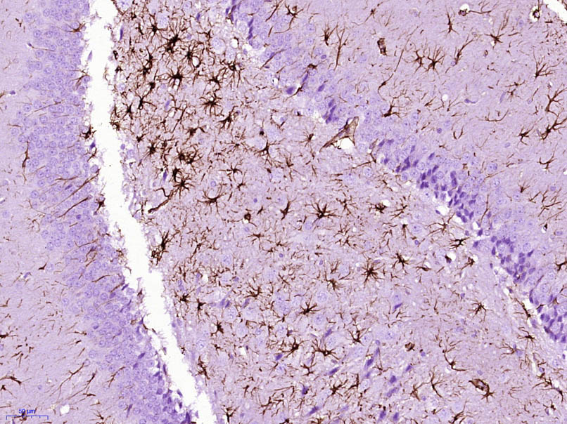 Paraformaldehyde-fixed, paraffin embedded (Rat brain); Antigen retrieval by boiling in sodium citrate buffer (pH6.0) for 15min; Block endogenous peroxidase by 3% hydrogen peroxide for 20 minutes; Blocking buffer (normal goat serum) at 37°C for 30min; Antibody incubation with (GFAP) Monoclonal Antibody, Unconjugated (bsm-33065M) at 1:400 overnight at 4°C, followed by operating according to SP Kit(Mouse) (sp-0024) instructionsand DAB staining. 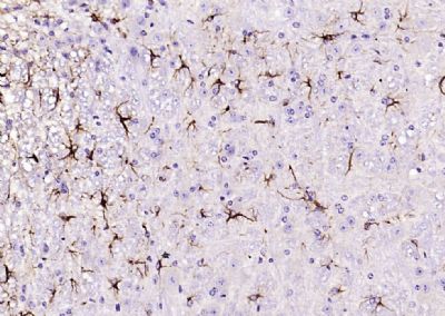 Paraformaldehyde-fixed, paraffin embedded (mouse cerebellum); Antigen retrieval by boiling in sodium citrate buffer (pH6.0) for 15min; Block endogenous peroxidase by 3% hydrogen peroxide for 20 minutes; Blocking buffer (normal goat serum) at 37°C for 30min; Antibody incubation with (GFAP) Monoclonal Antibody, Unconjugated (bsm-33065M) at 1:200 overnight at 4°C, followed by operating according to SP Kit(Mouse)(sp-0024) instructionsand DAB staining. 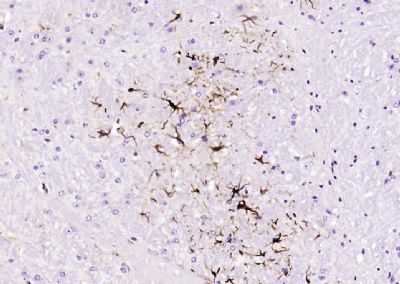 Paraformaldehyde-fixed, paraffin embedded (mouse cerebellum); Antigen retrieval by boiling in sodium citrate buffer (pH6.0) for 15min; Block endogenous peroxidase by 3% hydrogen peroxide for 20 minutes; Blocking buffer (normal goat serum) at 37°C for 30min; Antibody incubation with (GFAP) Monoclonal Antibody, Unconjugated (bsm-33065M) at 1:200 overnight at 4°C, followed by operating according to SP Kit(Mouse)(sp-0024) instructionsand DAB staining. 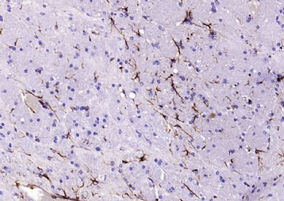 Paraformaldehyde-fixed, paraffin embedded (mouse brain); Antigen retrieval by boiling in sodium citrate buffer (pH6.0) for 15min; Block endogenous peroxidase by 3% hydrogen peroxide for 20 minutes; Blocking buffer (normal goat serum) at 37°C for 30min; Antibody incubation with (GFAP) Monoclonal Antibody, Unconjugated (bsm-33065M) at 1:200 overnight at 4°C, followed by operating according to SP Kit(Mouse)(sp-0024) instructionsand DAB staining. 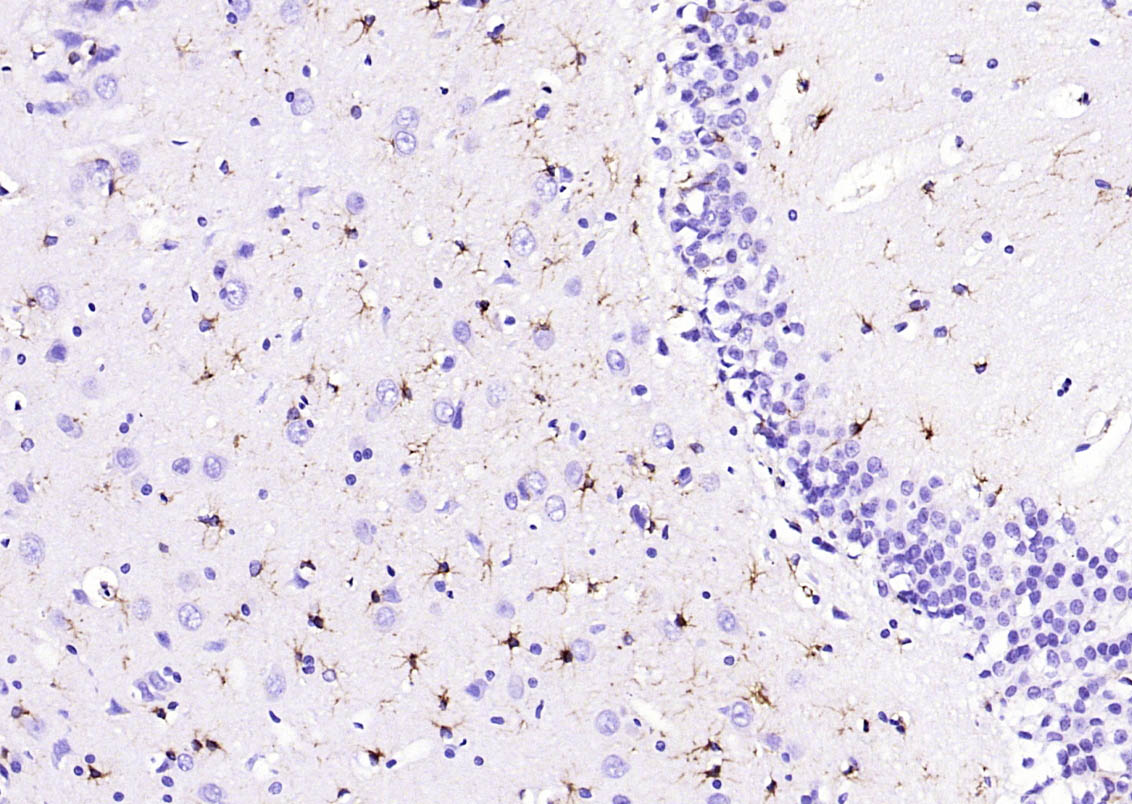 Paraformaldehyde-fixed, paraffin embedded (rat brain); Antigen retrieval by boiling in sodium citrate buffer (pH6.0) for 15min; Block endogenous peroxidase by 3% hydrogen peroxide for 20 minutes; Blocking buffer (normal goat serum) at 37°C for 30min; Antibody incubation with (GFAP) Monoclonal Antibody, Unconjugated (bsm-33065M) at 1:200 overnight at 4°C, followed by operating according to SP Kit(Mouse)(sp-0024) instructionsand DAB staining. 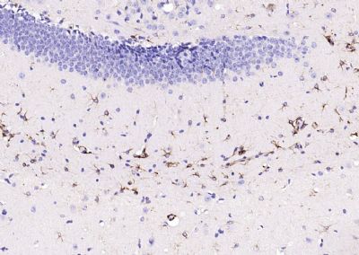 Paraformaldehyde-fixed, paraffin embedded (mouse brain); Antigen retrieval by boiling in sodium citrate buffer (pH6.0) for 15min; Block endogenous peroxidase by 3% hydrogen peroxide for 20 minutes; Blocking buffer (normal goat serum) at 37°C for 30min; Antibody incubation with (GFAP) Monoclonal Antibody, Unconjugated (bsm-33065M) at 1:200 overnight at 4°C, followed by operating according to SP Kit(Mouse)(sp-0024) instructionsand DAB staining.  Paraformaldehyde-fixed, paraffin embedded (Mouse cerebellum); Antigen retrieval by boiling in sodium citrate buffer (pH6.0) for 15min; Block endogenous peroxidase by 3% hydrogen peroxide for 20 minutes; Blocking buffer (normal goat serum) at 37°C for 30min; Antibody incubation with (GFAP) Monoclonal Antibody, Unconjugated (bsm-33065M) at 1:800 overnight at 4°C, followed by operating according to SP Kit(Mouse) (sp-0024) instructions and DAB staining. 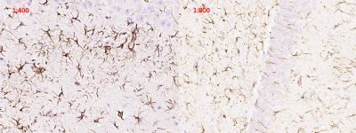 Paraformaldehyde-fixed, paraffin embedded (Rat brain); Antigen retrieval by boiling in sodium citrate buffer (pH6.0) for 15min; Block endogenous peroxidase by 3% hydrogen peroxide for 20 minutes; Blocking buffer (normal goat serum) at 37°C for 30min; Antibody incubation with (GFAP) Monoclonal Antibody, Unconjugated (bsm-33065M) at 1:400 and 1:800 overnight at 4°C, followed by operating according to SP Kit(Mouse) (sp-0024) instructions and DAB staining. 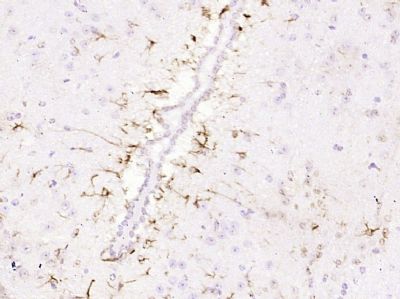 Paraformaldehyde-fixed, paraffin embedded (Mouse brain); Antigen retrieval by boiling in sodium citrate buffer (pH6.0) for 15min; Block endogenous peroxidase by 3% hydrogen peroxide for 20 minutes; Blocking buffer (normal goat serum) at 37°C for 30min; Antibody incubation with (GFAP) Monoclonal Antibody, Unconjugated (bsm-33065M) at 1:800 overnight at 4°C, followed by operating according to SP Kit(Mouse) (sp-0024) instructions and DAB staining. 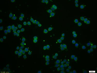 Tissue/cell: BV-2 cell; 4% Paraformaldehyde-fixed; Triton X-100 at room temperature for 20 min; Blocking buffer (normal goat serum, C-0005) at 37°C for 20 min; Antibody incubation with (GFAP) Monoclonal Antibody, Unconjugated (bsm-33065M) 1:50, 90 minutes at 37°C; followed by a conjugated Goat Anti-Mouse IgG antibody (bs-0296G-FITC) at 37°C for 90 minutes, DAPI (5ug/ml, blue, C-0033) was used to stain the cell nuclei. |
ĪŅŅŖŃƼŪ
*ĮŖĻµ·½Ź½£ŗ
(æÉŅŌŹĒQQ”¢MSN”¢µē×ÓÓŹĻ䔢µē»°µČ£¬ÄśµÄĮŖĻµ·½Ź½²»»į±»¹«æŖ)
*ÄŚČŻ£ŗ